. 2018 Jul 6:6:e5212.
doi: 10.7717/peerj.5212. eCollection 2018.

Xenoposeidon is the earliest known rebbachisaurid sauropod dinosaur

Affiliations

Xenoposeidon is the earliest known rebbachisaurid sauropod dinosaur

Michael P Taylor. PeerJ. .

Abstract

Xenoposeidon proneneukos is a sauropod dinosaur from the Early Cretaceous Hastings Group of England. It is represented by a single partial dorsal vertebra, NHMUK PV R2095, which consists of the centrum and the base of a tall neural arch. Despite its fragmentary nature, it is recognisably distinct from all other sauropods, and is here diagnosed with five unique characters. One character previously considered unique is here recognised as shared with the rebbachisaurid diplodocoid Rebbachisaurus garasbae from the mid-Cretaceous of Morocco: an 'M'-shaped arrangement of laminae on the lateral face of the neural arch. Following the more completely preserved R. garasbae, these laminae are now interpreted as ACPL and lateral CPRL, which intersect anteriorly; and PCDL and CPOL, which intersect posteriorly. Similar arrangements are also seen in some other rebbachisaurid specimens (though not all, possibly due to serial variation), but never in non-rebbachisaurid sauropods. Xenoposeidon is therefore referred to Rebbachisauridae. Due to its inferred elevated parapophysis, the holotype vertebra is considered a mid-posterior dorsal despite its elongate centrum. Since Xenoposeidon is from the Berriasian-Valanginian (earliest Cretaceous) Ashdown Formation of the Wealden Supergroup of southern England, it is the earliest known rebbachisaurid by some 10 million years. Electronic 3D models were invaluable in determining Xenoposeidon's true affinities: descriptions of complex bones such as sauropod vertebrae should always provide them where possible.

Keywords: 3D modelling; Dinosauria; Laminae; Rebbachisauridae; Sauropoda; Wealden; Xenoposeidon.


Conflict of interest statement

The authors declare that they have no competing interests.

Figures

Figure 1
Figure 1. NHMUK R2095, the holotype and only vertebra of Xenoposeidon proneneukos, shown from all six cardinal directions.
(A) Dorsal view, with anterior to the left. (B) anterior, (C) left lateral, (D) posterior, (E) right lateral view and (F) ventral view, with anterior to the left. Scale bar = 200 mm.
Figure 2
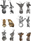
Figure 2. Comparative morphology of mid-posterior dorsals from six sauropods: Xenoposeidon and five representatives of major groups.
Each vertebra is shown in anterior and left lateral (or right lateral reversed) views, scaled to the same centrum height. (A) The diplodocid Diplodocus carnegii CM 84, 8th dorsal vertebra: (A1) anterior, modified from Hatcher (1901: plate VIII), (A2) right lateral reversed, modified from Hatcher (1901: plate VII). (B) The rebbachisaurid Rebbachisaurus garasbae MNHN MRS 1958, posterior dorsal vertebra: (B1) anterior, (B2) left lateral. (C) Xenoposeidon proneneukos NHMUK R2095, mid-posterior dorsal vertebra: (C1) anterior, (C2) left lateral. (D) The camarasaurid Camarasaurus supremus AMNH 5760/D-X-125, ?10th dorsal vertebra, modified from Osborn and Mook (1921: plate LXX): (D1) anterior, (D2) left lateral. (E) The brachiosaurid Giraffatitan brancai MB.R.3822 (formerly HMN AR1), from a digital model supplied by Heinrich Mallison: (E1) anterior, (E2) right lateral reversed. (F) The titanosaur Yongjinglong datangi GSGM ZH(08)-04, mid-dorsal vertebra, modified from Li et al. (2014: figure 9): (F1) anterior, (F2) left lateral.
Figure 3
Figure 3. Autapomorphies of Xenoposeidon proneneukos NHMUK R2095, mid-posterior dorsal vertebra, highlighted in red.
(A) Anterior view. (B) Left lateral view. Numbers pertain to the numbering of autapomorphies in the text. (1a), neural arch covers whole of centrum, and (1b) is contiguous with posterior articular facet. (2), neural arch is inclined forward by 35° relative to the vertical. (3a), inclined ridge-like lamina marks ventral margin of (3b) broad featureless area of bone. (4), large teardrop-shaped anterior fossa. (5a), vaulted laminae bound this fossa, but are not the medial CPRLs (5b, drawn in finer lines), which continue up to the presumed location of the prezygapophyses.
Figure 4
Figure 4. Centra and neural arches of posterior dorsal vertebrae from two rebbachisaurid sauropods (not to scale), highlighting the distinctive ‘M’ shape formed by laminae on the lateral face of the neural arch.
(A) NHMUK R2095, the holotype and only vertebra of Xenoposeidon proneneukos. (B) MNHN MRS 1958, a posterior dorsal vertebra from the holotype specimen of Rebbachisaurus garasbae.
Figure 5
Figure 5. NHMUK R2095, the holotype and only vertebra of Xenoposeidon proneneukos, in left anteroventrolateral view, highlighting the three sets of laminae related to the prezygapophyses.
The trajectories of the medial CPRLs (which emerge from the neural arch pedicels) and the lateral CPRLs (which intersect with the APCLs) indicate the approximate position of the prezygapophyses. The additional arched laminae form the margins of the large, teardrop-shaped anterior fossa, homologous with a CPRF, but meet at a position some way below and posterior to the presumed location of the prezygapophyseal facets. Breakage of both medial CPRLs and the left ACPL and PCDL is indicated by cross-hatching. Note that, from this perspective, the lateral CPRL appears to turn a corner where it intersects with the ACPL, such that the posteroventral portion of the lateral CPRL appears contiguous with the dorsal portion of the ACPL. This is an illusion brought about by the eminence at the point of intersection. As always, this is much easier to see in three dimensions (see File S1).
Figure 6
Figure 6. NHMUK R2095, the holotype and only vertebra of Xenoposeidon proneneukos, in left lateral view, with interpretative drawings.
(A) The incorrect interpretation of the laminae from Taylor & Naish (2007: figure 4A), with identifying captions greyed out since they are largely incorrect. (B) The revised interpretation of the same laminae, based on the similar arrangement in Rebbachisaurus garasbae. Scale bar = 200 mm.
Figure 7
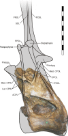
Figure 7. NHMUK R2095, the holotype and only vertebra of Xenoposeidon proneneukos, in left lateral view, interpreted as a rebbachisaurid.
This interpretation is modelled primarily on MNHN MRS 1958, a posterior dorsal vertebra from the holotype specimen of Rebbachisaurus garasbae. The CPOL passes through a sheetlike PCDL, as in Rebbachisaurus; but the lateral CPRL forms a cross-shaped junction with the ACPL, each of these laminae equally interrupting the trajectory of the other. Scale bar = 200 mm.

References

    1. Bonaparte JF. Les dinosaures (Carnosaures, Allosauridés, Sauropodes, Cétiosauridés) du Jurassique moyen de Cerro Cóndor (Chubut, Argentina) Annales de Paléontologie. 1986;72:325–386.
    1. Bonaparte JF. Evolucion de las vertebras presacras en Sauropodomorpha [Evolution of the presacral vertebrae in Sauropodomorpha] Ameghiniana. 1999;36(2):115–187.
    1. Borinder NH, Poropat SF, Kear BP. Reassessment of the earliest documented stegosaurian fossils from Asia. Cretaceous Research. 2016;68:61–69. doi: 10.1016/j.cretres.2016.08.004. - DOI
    1. Carballido JL, Rauhut OWM, Pol D, Salgado L. Osteology and phylogenetic relationships of Tehuelchesaurus benitezii (Dinosauria, Sauropoda) from the Upper Jurassic of Patagonia. Zoological Journal of the Linnean Society. 2011;163(2):605–662. doi: 10.1111/j.1096-3642.2011.00723.x. - DOI
    1. Chure D, Britt BB, Whitlock JA, Wilson JA. First complete sauropod dinosaur skull from the Cretaceous of the Americas and the evolution of sauropod dentition. Naturwissenschaften. 2010;97(4):379–391. doi: 10.1007/s00114-010-0650-6. - DOI - PMC - PubMed

LinkOut - more resources