

Regional cortical perfusion on arterial spin labeling MRI in dementia with Lewy bodies: Associations with clinical severity, glucose metabolism and tau PET
- PMID: 30003031
- PMCID: PMC6039836
- DOI: 10.1016/j.nicl.2018.06.020
Regional cortical perfusion on arterial spin labeling MRI in dementia with Lewy bodies: Associations with clinical severity, glucose metabolism and tau PET
Abstract
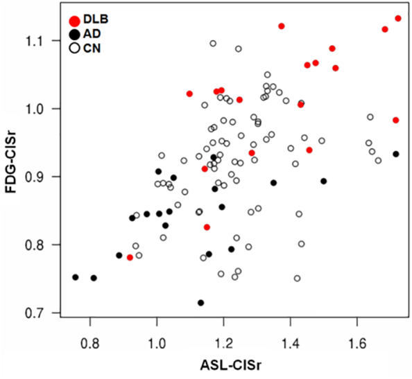
Visually preserved metabolism in posterior cingulate cortex relative to hypometabolism in precuneus and cuneus, the cingulate island sign, is a feature of dementia with Lewy bodies (DLB) on FDG-PET. Lower cingulate island sign ratio (posterior cingulate cortex/cuneus+precuneus; FDG-CISr) values have been associated with a higher Braak neurofibrillary tangle stage in autopsied DLB. Using voxel-wise analysis, we assessed the patterns of regional cortical perfusion and metabolism, and using an atlas-based approach, we measured perfusion cingulate island sign ratio on arterial spin labeling MRI (ASL-CISr), and its associations with FDG-CISr, uptake on tau-PET and clinical severity in DLB. Our study sample (n = 114) included clinically probable DLB patients (n = 19), age-matched patients with probable Alzheimer's disease dementia (AD; n = 19) and matched controls (n = 76) who underwent MRI with 3-dimensional pseudo-continuous arterial spin labeling, 18F-FDG-PET and 18F-AV-1451 tau PET. Patterns of cortical perfusion and metabolism were derived from quantitative maps using Statistical Parametric Mapping. DLB patients showed hypoperfusion on ASL-MRI in precuneus, cuneus and posterior parieto-occipital cortices, compared to controls, and relatively spared posterior cingulate gyrus, similar to pattern of hypometabolism on FDG-PET. DLB patients had higher ASL-CISr and FDG-CISr than AD patients (p <0.001). ASL-CISr correlated with FDG-CISr in DLB patients (r = 0.67; p =0.002). Accuracy of distinguishing DLB from AD patients was 0.80 for ASL-CISr and 0.91 for FDG-CISr. Lower ASL-CISr was moderately associated with a higher composite medial temporal AV-1451 uptake (r = -0.50; p =0.03) in DLB. Lower perfusion in precuneus and cuneus was associated with worse global clinical scores. In summary, the pattern of cortical hypoperfusion on ASL-MRI is similar to hypometabolism on FDG-PET, and respective cingulate island sign ratios correlate with each other in DLB. Non-invasive and radiotracer-free ASL-MRI may be further developed as a tool for the screening and diagnostic evaluation of DLB patients in a variety of clinical settings where FDG-PET is not accessible.
Keywords: Arterial spin labeling MRI; Cingulate island sign ratio; Cortical perfusion; Dementia with Lewy bodies; FDG PET; Or AV1–1451 tau PET.
Figures


References
-
- AASM . American Academy of Sleep Medicine; Chicago: 2005. International Classification of Sleep Disorders—2: Diagnostic and Coding Manual.
-
- Alsop D.C., Detre J.A., Golay X., Gunther M., Hendrikse J., Hernandez-Garcia L. Recommended implementation of arterial spin-labeled perfusion MRI for clinical applications: a consensus of the ISMRM perfusion study group and the European consortium for ASL in dementia. Magn. Reson. Med. 2015;73(1):102–116. - PMC - PubMed
-
- Binnewijzend M.A., Kuijer J.P., van der Flier W.M., Benedictus M.R., Moller C.M., Pijnenburg Y.A. Distinct perfusion patterns in Alzheimer's disease, frontotemporal dementia and dementia with Lewy bodies. Eur. Radiol. 2014;24(9):2326–2333. - PubMed
-
- Buxton R.B., Frank L.R. A model for the coupling between cerebral blood flow and oxygen metabolism during neural stimulation. J. Cereb. Blood Flow Metab. 1997;17(1):64–72. - PubMed
Publication types
MeSH terms
Substances
Grants and funding
LinkOut - more resources
Full Text Sources
Other Literature Sources
Medical
