. 2018 May 30:2018:6705842.
doi: 10.1155/2018/6705842. eCollection 2018.

Human MLL-AF9 Overexpression Induces Aberrant Hematopoietic Expansion in Zebrafish

Affiliations

Human MLL-AF9 Overexpression Induces Aberrant Hematopoietic Expansion in Zebrafish

Jiaqi Tan et al. Biomed Res Int. .

Erratum in

Abstract

The 11q23 of the mixed lineage leukemia 1 (MLL1) gene plays a crucial role in early embryonic development and hematopoiesis. The MLL-AF9 fusion gene, resulting from chromosomal translocation, often leads to acute myeloid leukemia with poor prognosis. Here, we generated a zebrafish model expressing the human MLL-AF9 fusion gene. Microinjection of human MLL-AF9 mRNA into zebrafish embryos resulted in enhanced hematopoiesis and the activation of downstream genes such as meis1 and hox cluster genes. Embryonic MLL-AF9 expression upregulated HSPC and myeloid lineage markers. Doxorubicin and MI-2 (a menin inhibitor) treatments significantly restored normal hematopoiesis in MLL-AF9-expressing animals. This study provides insight into the role of MLL-AF9 in zebrafish hematopoiesis and establishes a robust and efficient in vivo model for high-throughput drug screening.


Figures

Figure 1
Figure 1
MLL-AF9 overexpression in zebrafish embryos and activation of MLL downstream genes. MLL-AF9 mRNA was injected into zebrafish at the 1–2-cell stage (around 5ng per embryo). Injected embryos and control embryos were harvested at 48hpf. (a) Western blotting of the N-terminus of the MLL protein in MLL-AF9-injected and control embryos. (b) Relative expression of MLL downstream genes analyzed by qRT-PCR. (c–e) WISH assays of hoxa9a, hoxb5a, and meis1 in MLL-AF9-injected and control embryos at 24hpf. (f, g) Morphology of the hematopoietic cells of control embryos and MLL-AF9-injected embryos at 48 hpf.
Figure 2
Figure 2
Induction of zebrafish primitive and definitive hematopoiesis by MLL-AF9. WISH assays and qRT-PCR analysis of hematopoietic markers in MLL-AF9-injected and control embryos. (a–c) WISH assays of hematopoietic markers (lmo2 and gata1) and a vasculature formation marker (fli) in MLL-AF9-injected and control embryos at 24hpf. (d–h) WISH assays at 48hpf of the definitive hematopoietic stem cell marker c-myb, the erythropoietic marker hbbe1, and the myeloid lineage markers l-plastin, mpy, and lyz. Relative expression of the marker genes for (i) primitive hematopoiesis at 24hpf, (j) hematopoiesis at 48hpf, (k) vasculature formation (flk, fli) at 24hpf, and (k) definitive hematopoiesis initiation (c-myb and runx1) at 48hpf analyzed by qRT-PCR.
Figure 3
Figure 3
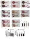
Doxorubicin inhibition of MLL-AF9-induced hematopoiesis. Doxorubicin and DMSO (control) were administered to MLL-AF9-injected and wild-type embryos from 24 to 48hpf. WISH assays of the (a) definitive hematopoietic stem cell marker c-myb and (b–d) myeloid lineage markers mpo, lyz, and l-plastin in wild-type embryos treated with DMSO (top), MLL-AF9-injected embryos treated with DMSO (middle), and MLL-AF9-injected embryos treated with doxorubicin (bottom) at 48hpf. (e) Relative expression of the marker genes for definitive hematopoiesis (runx1 and c-myb) and myeloid hematopoiesis (l-plastin, csf1r, mfap4, mpo, and lyz) in wild-type and MLL-AF9-injected embryos treated with DMSO or doxorubicin analyzed by qRT-PCR. Wild-type embryos treated with DMSO were used as a control.
Figure 4
Figure 4
Amelioration of MLL-AF9-induced hematopoiesis by MI-2. The menin inhibitor MI-2 (2.5mmol/L) and DMSO (control) were administered to MLL-AF9-injected and wild-type embryos from 24 to 72hpf. WISH assays of the (a) definitive hematopoietic stem cell marker c-myb and (b–d) myeloid lineage markers mpo, lyz, and l-plastin in wild-type embryos treated with DMSO (top), MLL-AF9-injected embryos treated with DMSO (middle), and MLL-AF9-injected embryos treated with MI-2 (bottom) at 72hpf. (e) Relative expression of the marker genes for definitive hematopoiesis (runx1 and c-myb) and myeloid hematopoiesis (l-plastin, csf1r, mfap4, mpo, and lyz) in wild-type and MLL-AF9-injected embryos treated with MI-2 or doxorubicin analyzed by qRT-PCR. Wild-type embryos treated with DMSO were used as a control.

Similar articles

Cited by

References

    1. Marschalek R. Systematic classification of mixed-lineage leukemia fusion partners predicts additional cancer pathways. Annals of Laboratory Medicine. 2016;36(2):85–100. doi: 10.3343/alm.2016.36.2.85. - DOI - PMC - PubMed
    1. Tamai H., Inokuchi K. 11q23/MLL acute leukemia: update of clinical aspects. Journal of Clinical and Experimental Hematopathology. 2010;50(2):91–98. doi: 10.3960/jslrt.50.91. - DOI - PubMed
    1. Marschalek R. MLL leukemia and future treatment strategies. Das Archiv der Pharmazie: Chemistry in Life Sciences. 2015;348(4):221–228. doi: 10.1002/ardp.201400449. - DOI - PubMed
    1. de Boer J., Walf-Vorderwülbecke V., Williams O. In focus: MLL-rearranged leukemia. Leukemia. 2013;27(6):1224–1228. doi: 10.1038/leu.2013.78. - DOI - PubMed
    1. Yu B. D., Hanson R. D., Hess J. L., Horning S. E., Korsmeyer S. J. MLL, a mammalian trithorax-group gene, functions as a transcriptional maintenance factor in morphogenesis. Proceedings of the National Acadamy of Sciences of the United States of America. 1998;95(18):10632–10636. doi: 10.1073/pnas.95.18.10632. - DOI - PMC - PubMed

Substances

LinkOut - more resources