. 2018 Oct;17(5):e12815.
doi: 10.1111/acel.12815. Epub 2018 Jul 12.

In vivo GDF3 administration abrogates aging related muscle regeneration delay following acute sterile injury

Affiliations

In vivo GDF3 administration abrogates aging related muscle regeneration delay following acute sterile injury

Andreas Patsalos et al. Aging Cell. 2018 Oct.

Abstract

Tissue regeneration is a highly coordinated process with sequential events including immune cell infiltration, clearance of damaged tissues, and immune-supported regrowth of the tissue. Aging has a well-documented negative impact on this process globally; however, whether changes in immune cells per se are contributing to the decline in the body's ability to regenerate tissues with aging is not clearly understood. Here, we set out to characterize the dynamics of macrophage infiltration and their functional contribution to muscle regeneration by comparing young and aged animals upon acute sterile injury. Injured muscle of old mice showed markedly elevated number of macrophages, with a predominance for Ly6Chigh pro-inflammatory macrophages and a lower ratio of the Ly6Clow repair macrophages. Of interest, a recently identified repair macrophage-derived cytokine, growth differentiation factor 3 (GDF3), was markedly downregulated in injured muscle of old relative to young mice. Supplementation of recombinant GDF3 in aged mice ameliorated the inefficient regenerative response. Together, these results uncover a deficiency in the quantity and quality of infiltrating macrophages during aging and suggest that in vivo administration of GDF3 could be an effective therapeutic approach.


Figures

Figure 1
Figure 1
Impaired skeletal muscle regeneration and delayed phenotypic transition of infiltrating myeloid cells in aged animals following CTX injury. (a) Representative images of H&E‐stained skeletal muscle from young adult (2‐month‐old) and aged (23‐month‐old) male mice at Days 0, 8, 12, and 16 post‐CTX‐induced injury. Scale bars in the upper left represent 100 μm. (b) Mean myofiber cross‐sectional area (CSA) of regenerating muscles in young adult (2‐month‐old) and aged (23‐month‐old) mice (number of fibers counted > 20,000) at Days 0, 8, 12, and 16 post‐CTX‐induced injury (n = 6 per group). (c) The ratio of necrotic fibers relative to regeneration area (in mm2) at Day 8 of regeneration in young adult (2‐month‐old) and aged (23‐month‐old) muscle sections is shown. (d) Normalized tibialis anterior (TA) muscle mass‐to‐body weight ratio from young adult (2‐month‐old) and aged (23‐month‐old) mice at indicated time points following CTX injury (n = 6 per group). (e) Number of infiltrating myeloid (CD45+) cells in regenerating muscle from young (2‐month‐old) or aged (28‐month‐old) muscles at indicated time points prior and post‐CTX injury (n = 8 muscles per group). (f) Heatmap representations of atrophy and macrophage‐related genes (measured by qPCR) from young and old uninjured (left panel) and regenerating (Day 8 post‐CTX; right panel) TA muscles. Relative mRNA expression (calculated using the 2‐ΔΔCT method) is shown as log10(fold change) (n = 6 muscles per group). (g and h) Percentage of inflammatory (Ly6Chigh F4/80low) and repair (Ly6Clow F4/80high) MFs from young (2‐month‐old) or aged (28‐month‐old) muscles at indicated time points following CTX injury (n = 8 mice per group). In all bar and line graphs, bars and data points represent mean ± SEM
Figure 2
Figure 2
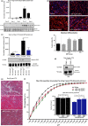
GDF3 induces myoblast differentiation and improves the kinetics of regeneration in aged animals. (a) GDF3 protein expression in whole‐muscle lysates of regenerating TA muscles from young adult male mice (2‐month‐old) at indicated time points (D, day) post‐CTX injury. Three representative biological replicates are shown for each time point (lower panel), and mean normalized signal values to total protein of each group are shown (upper panel) (n = 6). REVERT total protein was used for loading control and signal normalization. (b) Immunofluorescence against desmin (red) and DAPI (blue) shows a drastic enhancement of myotube formation in the presence of recombinant (r) GDF3 in primary myoblasts (n = 4). Representative images from 100, 300, and 600 ng/ml rGDF3‐treated myoblasts are shown (upper panel). Lower panel shows the fusion index of primary myoblasts in the presence of various concentrations of recombinant GDF3 (n = 4). Two different sources of the protein were used. SBP stands for the in‐house Sanford Burnham Prebys Protein Core produced version and R&D for the commercially available one. (c) Decreased GDF3 protein expression in whole‐muscle lysates of regenerating muscles from aged (28‐month‐old) mice at different time points (D, day). Two representative biological replicates are shown for each group (lower panel) with mean normalized signal values to total protein of each group shown in the upper panel (n = 6). REVERT total protein was used for loading control and signal normalization. (d) GDF3 protein expression in CD45+ and CD45 cells isolated at Day 4 post‐CTX injury from aged (23‐month‐old) mice. REVERT total protein was used for loading control. (e, f) Improvement in regeneration by administration of recombinant GDF3 (300 and 600 ng) in young (2‐month‐old) or aged (28‐month‐old) male animals (n = 6 per group). (e) H&E‐stained images from aged treated mice, and (f) cumulated and mean CSA (right panel) measurements are shown. In all bar graphs, bars represent mean ± SEM

References

    1. Bernet, J. D. , Doles, J. D. , Hall, J. K. , Kelly Tanaka, K. , Carter, T. A. , & Olwin, B. B. (2014). p38 MAPK signaling underlies a cell‐autonomous loss of stem cell self‐renewal in skeletal muscle of aged mice. Nature Medicine, 20(3), 265–271. 10.1038/nm.3465 - DOI - PMC - PubMed
    1. Brack, A. S. , Conboy, M. J. , Roy, S. , Lee, M. , Kuo, C. J. , Keller, C. , & Rando, T. A. (2007). Increased Wnt signaling during aging alters muscle stem cell fate and increases fibrosis. Science, 317(5839), 807–810. 10.1126/science.1144090 - DOI - PubMed
    1. Chakkalakal, J. V. , Jones, K. M. , Basson, M. A. , & Brack, A. S. (2012). The aged niche disrupts muscle stem cell quiescence. Nature, 490(7420), 355–360. 10.1038/nature11438 - DOI - PMC - PubMed
    1. Charge, S. B. , Brack, A. S. , & Hughes, S. M. (2002). Aging‐related satellite cell differentiation defect occurs prematurely after Ski‐induced muscle hypertrophy. American Journal of Physiology. Cell Physiology, 283(4), C1228–C1241. 10.1152/ajpcell.00206.2002 - DOI - PubMed
    1. Cheung, T. H. , & Rando, T. A. (2013). Molecular regulation of stem cell quiescence. Nature Reviews Molecular Cell Biology, 14(6), 329–340. 10.1038/nrm3591 - DOI - PMC - PubMed

Substances