Review
. 2018 Jul;6(4):10.1128/microbiolspec.rwr-0020-2018.
doi: 10.1128/microbiolspec.RWR-0020-2018.

Genes within Genes in Bacterial Genomes

Affiliations
Review

Genes within Genes in Bacterial Genomes

Sezen Meydan et al. Microbiol Spectr. 2018 Jul.

Abstract

Genetic coding in bacteria largely operates via the "one gene-one protein" paradigm. However, the peculiarities of the mRNA structure, the versatility of the genetic code, and the dynamic nature of translation sometimes allow organisms to deviate from the standard rules of protein encoding. Bacteria can use several unorthodox modes of translation to express more than one protein from a single mRNA cistron. One such alternative path is the use of additional translation initiation sites within the gene. Proteins whose translation is initiated at different start sites within the same reading frame will differ in their N termini but will have identical C-terminal segments. On the other hand, alternative initiation of translation in a register different from the frame dictated by the primary start codon will yield a protein whose sequence is entirely different from the one encoded in the main frame. The use of internal mRNA codons as translation start sites is controlled by the nucleotide sequence and the mRNA folding. The proteins of the alternative proteome generated via the "genes-within-genes" strategy may carry important functions. In this review, we summarize the currently known examples of bacterial genes encoding more than one protein due to the utilization of additional translation start sites and discuss the known or proposed functions of the alternative polypeptides in relation to the main protein product of the gene. We also discuss recent proteome- and genome-wide approaches that will allow the discovery of novel translation initiation sites in a systematic fashion.


Figures

FIGURE 1
FIGURE 1
Different strategies for encoding more than one protein in one gene. (a) The conventional one gene-one protein scenario. (b) Programmed ribosomal frameshifting results in translation of two proteins whose N-terminal sequences are the same but differ in their C-terminal segments. The alternative protein could be shorter or longer than the primary one depending on the position of the OOF stop codon. (c) Presence of an internal in-frame start codon within the ORF results in production of a truncated protein devoid of the N-terminal segment of the full-size translation product. If the internal start codon is OOF relative to the main ORF, the sequence of the alternative polypeptide is completely different from that of the protein encoded in the main ORF.
FIGURE 2
FIGURE 2
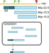
Internal initiation alters protein localization. Full-size Mip-23.5 protein carries a signal sequence (SS) at its N terminus, which predisposes it to be secreted. The products of internal initiation, Mip-15.5 and Mip-15.0, lack the signal sequence and, as a result, remain in the cytoplasm.
FIGURE 3
FIGURE 3
Alternative translation initiation accounts for production of two antibiotics by the same biochemical pathway. Production of the full-size PikAIV polyketide synthase module supports synthesis of the 14-atom macrolactone ring of pikromycin. The N-terminally truncated PikAIV variant leads to the synthesis of the smaller (12-atom) macrolactone ring of methymycin.
FIGURE 4
FIGURE 4
Alternative protein restores stoichiometry of the functional restriction enzyme. Full-size McrBL binds DNA and associates with the nucleolytic McrC to cleave DNA. McrBS lacks the N-terminal DNA-binding domain (dark blue) but is able to titrate out the excess McrC to maintain optimal activity of the restriction enzyme complex. The general scheme of the figure was adapted from reference .
FIGURE 5
FIGURE 5
The isoforms of CcmM help to differentially organize RubisCO in β-carboxysome. The N-terminal segment of the full-size CcmM-58 (dark blue) anchors it to the inner shell of the β-carboxysome, while its C-terminal segment (light blue) arranges the first layer of RubisCO. The N-terminally truncated Ccm-35 organizes RubisCO into a lattice in the lumen of the β-carboxysome.
FIGURE 6
FIGURE 6
The alternative LysC isoform expands the amino acid-mediated regulation of the enzyme. The full-size (α) and N-terminally truncated (β) isoforms of LysC associate into a functional α2β2 tetramer, in which the binding sites for the regulatory amino acids lysine (orange) and threonine (red) are formed at the interface of the α-β subunits. Shown is the structure of Corynebacterium glutamicum LysC (PDB ID: 3AAW) (95).
FIGURE 7
FIGURE 7
Elimination of the stop codon creates a fused gene with two translation starts. Two genes, tilS and hprT, which encode functionally distinct proteins and associate to form a functional complex, are organized in an operon in B. subtilis. Due to the elimination of the tilS stop codon in L. monocytogenes, the two ORFs are fused into a single gene and can be expressed from the primary and the internal TIS.
FIGURE 8
FIGURE 8
The use of different promoters can regulate the utilization of alternative start sites within a gene. Two promoters (P1 and P2) precede the petH gene in Synechocystis 6803. The transcript initiated at P2 has a short 5′ UTR; the accessible pTIS favors translation of the full-size FNRL. Folding of the long 5′ UTR of the transcripts initiated at the P1 promoter occludes the pTIS, thereby shifting the relative expression of the FNR isoforms in favor of the shorter FNRS. While photosynthetic growth favors synthesis of FNRL, heterotrophic conditions stimulate production of the FNRS isoform.
FIGURE 9
FIGURE 9
Elimination of a start codon leads to gene occlusion. In E. coli, the neighboring genes of ribosomal protein L34 (rpmH) and RNase P protein (rnpA) are independently translated. In T. thermophilus, the mutation of the rnpA translation initiation codon and appearance of an additional start site upstream of rpmH lead to occlusion of rpmH within the rnpA gene, but in an alternative reading frame. Start sites of both genes utilize the same SD sequence (blue), whose optimal spacing with the rpmH start codon shifts the balance of translation in favor of the ribosomal protein.
FIGURE 10
FIGURE 10
Retapamulin-assisted ribosome profiling illuminates sites of internal translation initiation. (a) (Left) Ribosome profiling in untreated cells shows the distribution of translating ribosomes along the mRNAs (30). (Right) Brief pretreatment of cells with the translation initiation inhibitor retapamulin arrests the ribosomes at the translation start site of the ORFs. (b) Examples of primary and internal TISs revealed by retapamulin-assisted ribosome profiling in E. coli genes known to contain functional internal start codons. Note that the presence of the iTISs is difficult to detect when ribosome profiling is performed in untreated cells.

References

    1. Atkins JF, Loughran G, Bhatt PR, Firth AE, Baranov PV. 2016. Ribosomal frameshifting and transcriptional slippage: from genetic steganography and cryptography to adventitious use. Nucleic Acids Res 44:7007–7078. - PMC - PubMed
    1. Baranov PV, Atkins JF, Yordanova MM. 2015. Augmented genetic decoding: global, local and temporal alterations of decoding processes and codon meaning. Nat Rev Genet 16:517–529. 10.1038/nrg3963. - DOI - PubMed
    1. Caliskan N, Peske F, Rodnina MV. 2015. Changed in translation: mRNA recoding by -1 programmed ribosomal frameshifting. Trends Biochem Sci 40:265–274. 10.1016/j.tibs.2015.03.006. - DOI - PMC - PubMed
    1. Dinman JD. 2012. Mechanisms and implications of programmed translational frameshifting. Wiley Interdiscip Rev RNA 3:661–673 10.1002/wrna.1126. - DOI - PMC - PubMed
    1. Miller ES, Kutter E, Mosig G, Arisaka F, Kunisawa T, Rüger W. 2003. Bacteriophage T4 genome. Microbiol Mol Biol Rev 67:86–156. 10.1128/MMBR.67.1.86-156.2003. [PubMed] - DOI - PMC - PubMed

Publication types

LinkOut - more resources