. 2018 Sep;62(17):e1800341.
doi: 10.1002/mnfr.201800341. Epub 2018 Jul 29.

Glycation of the Major Milk Allergen β-Lactoglobulin Changes Its Allergenicity by Alterations in Cellular Uptake and Degradation

Affiliations

Glycation of the Major Milk Allergen β-Lactoglobulin Changes Its Allergenicity by Alterations in Cellular Uptake and Degradation

Marija Perusko et al. Mol Nutr Food Res. 2018 Sep.

Abstract

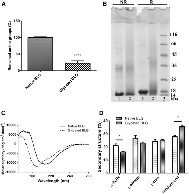
Scope: During food processing, the Maillard reaction (МR) may occur, resulting in the formation of glycated proteins. Glycated proteins are of particular importance in food allergies because glycation may influence interactions with the immune system. This study compared native and extensively glycated milk allergen β-lactoglobulin (BLG), in their interactions with cells crucially involved in allergy.

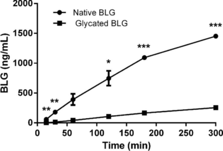
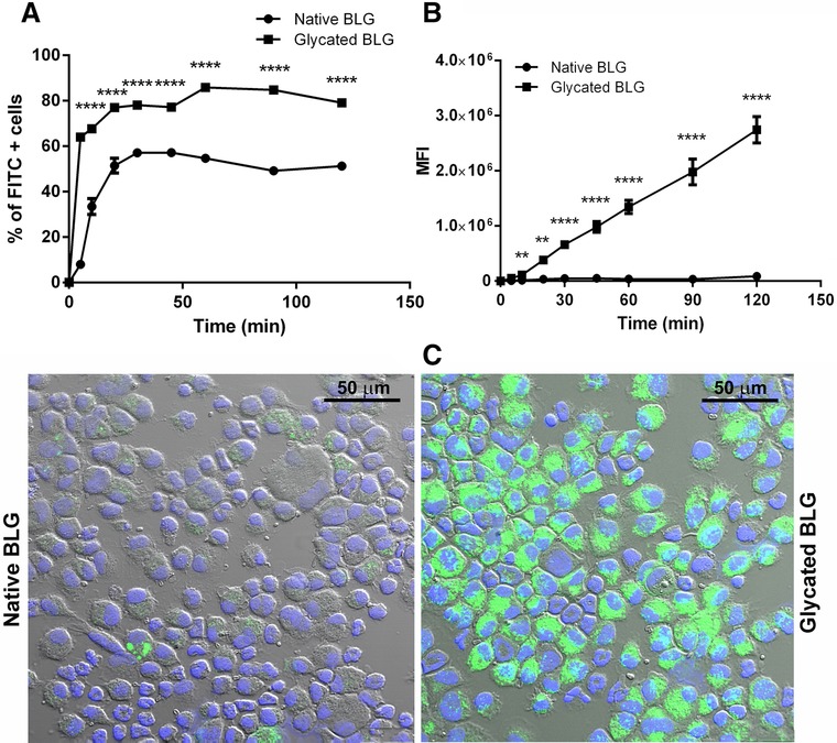
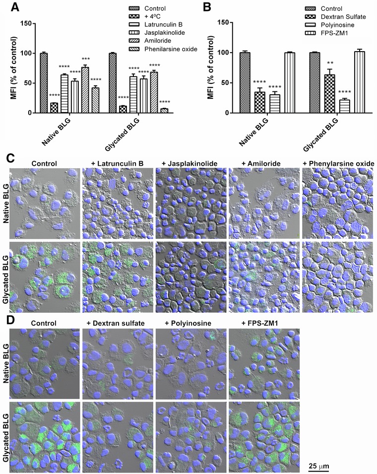
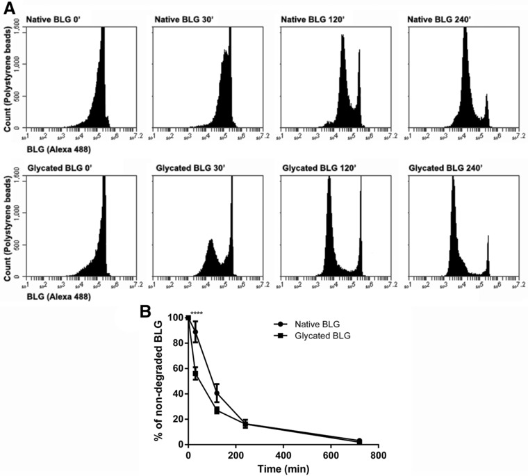
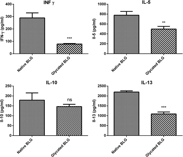
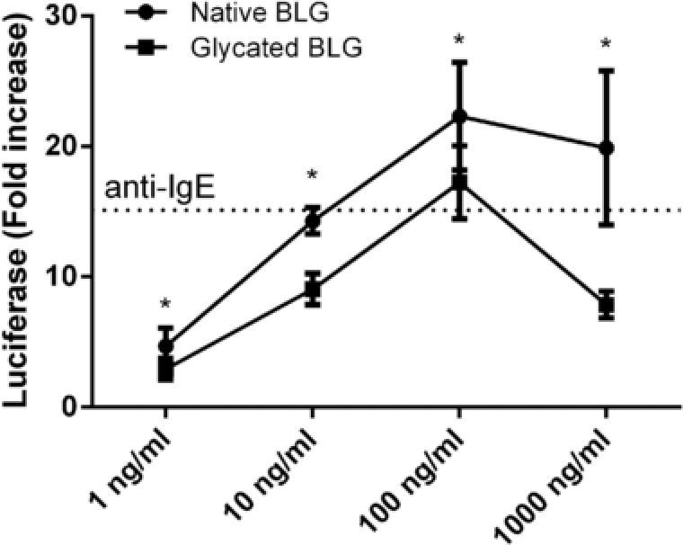
Methods and results: BLG was glycated in MR and characterized. Native and glycated BLG were tested in experiments of epithelial transport, uptake and degradation by DCs, T-cell cytokine responses, and basophil cell degranulation using ELISA and flow cytometry. Glycation of BLG induced partial unfolding and reduced its intestinal epithelial transfer over a Caco-2 monolayer. Uptake of glycated BLG by bone marrow-derived dendritic cells (BMDC) was increased, although both BLG forms entered BMDC via the same mechanism, receptor-mediated endocytosis. Once inside the BMDC, glycated BLG was degraded faster, which might have led to observed lower cytokine production in BMDC/CD4+ T-cells coculture. Finally, glycated BLG was less efficient in induction of degranulation of BLG-specific IgE sensitized basophil cells.

Conclusions: This study suggests that glycation of BLG by MR significantly alters its fate in processes involved in immunogenicity and allergenicity, pointing out the importance of food processing in food allergy.

Keywords: Maillard reaction; food allergens; food processing; uptake and degradation by DCs; β-lactoglobulin.


Figures

Figure 1
Figure 1
Characterization of BLG glycated in MR. A) Estimation of the degree of MR by determining remaining free amino group content by OPA method. Free amino group content was statistically compared by Student's t‐test. ****Denotes significance at p < 0.0001 confidence level. B) Protein profile of native and glycated BLG on 14% SDS‐PAGE gel under nonreducing (NR) and reducing (R) conditions. Protein bands were stained with CBB. 1) native BLG; 2) glycated BLG; 3) molecular weight markers (kDa). C) CD spectra of native and glycated BLG in far‐UV spectral range. Samples were recorded in 50 mm sodium phosphate buffer, pH 8.0 at protein concentration 0.85 mg mL. D) Percentages of the secondary structures were estimated by CONTIN algorithm available in CDPro package based on SP29 reference set. Secondary structure fractions were compared by Student's t‐test. *Represents significance at p < 0.05 confidence level.
Figure 2
Figure 2
Bioavailability of native and glycated BLG in vitro. Transcytosis of native and glycated BLG across Caco‐2 monolayer. Transport was measured by determining BLG concentration in samples withdrawn from basolateral chamber at different time‐points up to 3 h. Each point represents mean ± SD. One representative experiment out of three independent is shown. *, **, and *** represent significance at p < 0.05, 0.01, and 0.001, respectively.
Figure 3
Figure 3
BMDCs uptake of FITC‐labeled native and glycated BLG followed in time. BMDCs were incubated with 10 μg mL−1 FITC‐labeled native and glycated BLG for 120 min and the BMDC uptake was analyzed by means of flow cytometry. Data are expressed as A) percent of FITC‐positive BMDCs and B) MFI. ** and **** represent significance at p < 0.01 and 0.0001, respectively. C) Confocal micrographs of BMDC incubated with FITC‐labeled native and glycated BLG for 30 min.
Figure 4
Figure 4
Mechanistic studies of uptake of FITC‐labeled native and glycated BLG in the presence of different inhibitors. BMDCs were pre‐incubated for 30 min with A) mechanism‐specific inhibitors, namely: latrunculin B—inhibitor of actin polymerization; jasplackinolide—inhibitor of phagocytosis; amiloride—macropinocytosis inhibitor; phenylarsine oxide—inhibitor of receptor‐mediated endocytosis, or with B) receptor‐specific inhibitors: dextran sulfate and polyinosine—SRs inhibitors; FPS‐ZM1—RAGE inhibitor. Subsequently, cells were loaded with 10 μg mL−1 FITC‐labeled native and glycated BLG. BMDCs uptake was analyzed by flow cytometry, and data are represented as percent of MFI of non‐inhibited uptake as mean ± SEM (n = 6) and analyzed by Student's t‐tests. **, ***, and **** represent significance at p < 0.01, 0.001, and 0.0001, respectively. Mechanistic studies in the presence of C) mechanism‐specific inhibitors or D) receptor‐specific inhibitors were additionally followed by confocal microscopy.
Figure 5
Figure 5
Kinetics of endolysosomal degradation of native and glycated BLG. BMDC were allowed to internalize native and glycated BLG‐coated polystyrene beads and incubated for different periods at 37 °C to allow endolysosomal degradation. After every time‐point, cells were lysed and beads carrying non‐degraded BLG were stained by Alexa 488 BLG‐specific antibodies. Polystyrene beads were analyzed by flow cytometry. A) Flow cytometry dot plot of one representative experiment. B) Percentage of non‐degraded native and glycated BLG over time is shown as mean ± SEM (n = 4). **** denotes significance at p < 0.0001 confidence level.
Figure 6
Figure 6
Stimulation of BLG‐specific CD4+ T‐cells by native or glycated BLG‐primed BMDCs. CD4+ T‐cells isolated from C3H/HeOuJ mice immunized with native BLG were cocultured with BMDCs primed with native or glycated BLG for 72 h. Levels of INF‐γ, IL‐5, IL‐10, and IL‐13 in coculture supernatants were measured by ELISA. The data are presented as mean ± SD of one representative experiment and analyzed by Student's t‐test. **, ***, and ns, represent p < 0.01, 0.001, and not significant, respectively.
Figure 7
Figure 7
IgE crosslinking‐induced luciferase expression assay demonstrating basophil activation by native and glycated BLG. RS‐ATL8 cells were sensitized with the pool of BLG‐specific chimeric human IgE monoclonal antibodies and subsequently stimulated with 1, 10, 100, or 1000 ng mL−1 of native and glycated BLG. Luciferase expression level after 1 h of stimulation is shown. Dashed line shows luciferase expression after stimulation with 1 μg mL−1 anti‐human IgE antibodies. Data are expressed as mean ± SD (n = 8) and analyzed by t‐tests. *p < 0.05.

References

    1. Henle T., Amino Acids 2005, 29, 313. - PubMed
    1. Spotti M. J., Perduca M. J., Piagentini A., Santiago L. G., Rubiolo A. C., Carrara C. R., Food Hydrocoll. 2013, 31, 26.
    1. Zhu D., Damodaran S., Lucey J. A., J. Agric. Food Chem. 2010, 58, 2988. - PubMed
    1. Herceg Z., Režek A., Lelas V., Krešić G., Franetović M., J. Food Eng. 2007, 79, 279.
    1. Liu G., Zhong Q., J. Agric. Food Chem. 2012, 60, 9754. - PubMed

Publication types

MeSH terms