Comparative Study
. 2018 Oct 1;315(4):H934-H949.
doi: 10.1152/ajpheart.00238.2018. Epub 2018 Jul 13.

Characterization of a mouse model of obesity-related fibrotic cardiomyopathy that recapitulates features of human heart failure with preserved ejection fraction

Affiliations
Comparative Study

Characterization of a mouse model of obesity-related fibrotic cardiomyopathy that recapitulates features of human heart failure with preserved ejection fraction

Linda Alex et al. Am J Physiol Heart Circ Physiol. .

Abstract

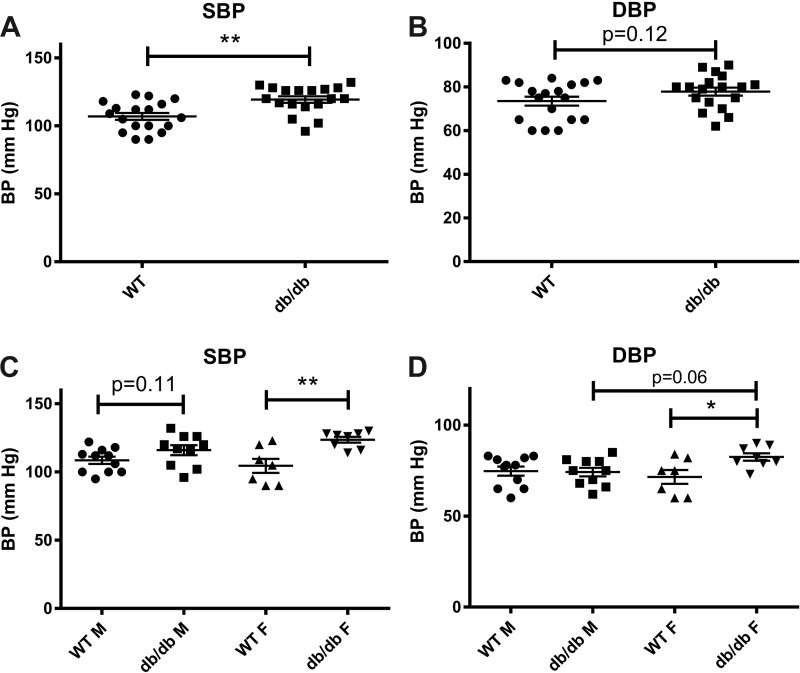
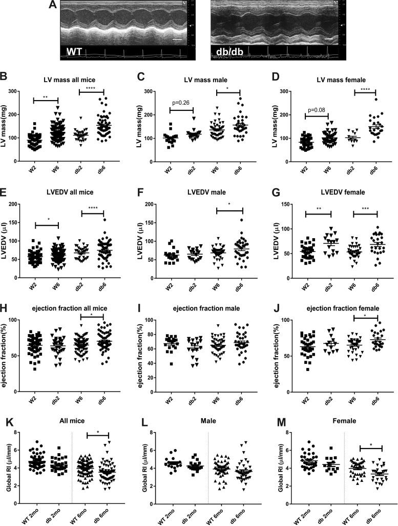
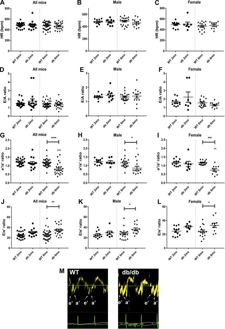
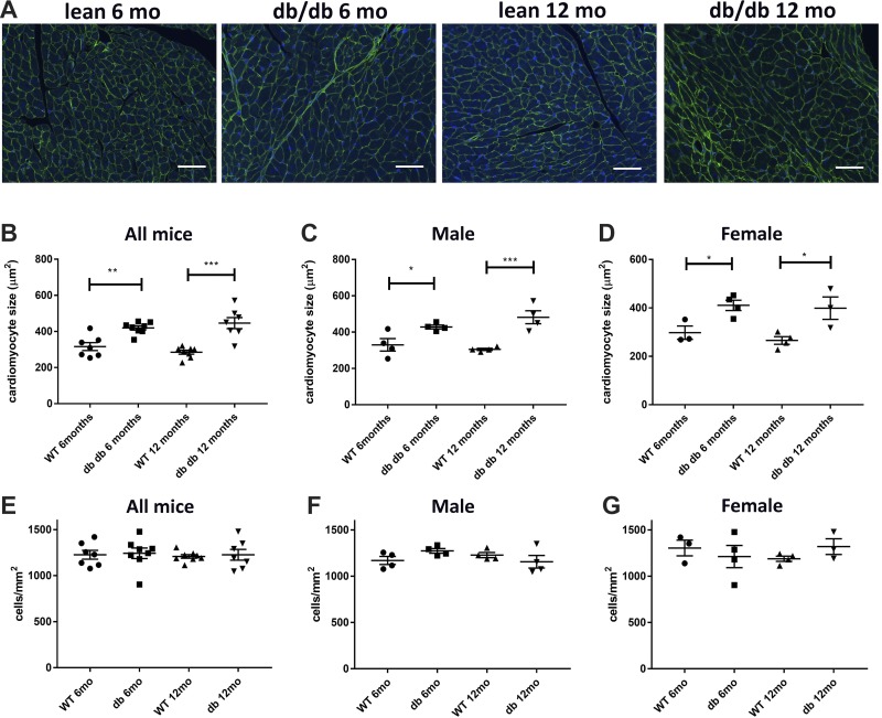
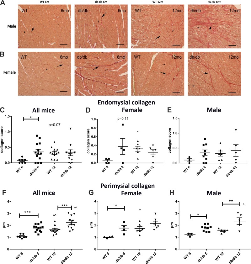
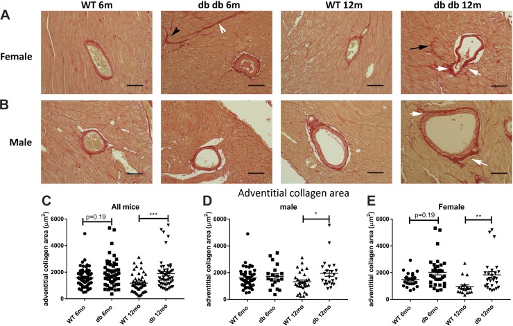
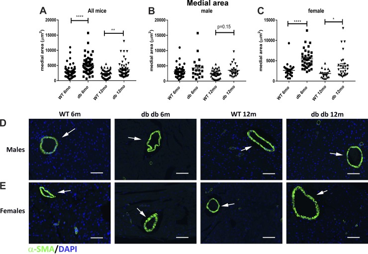
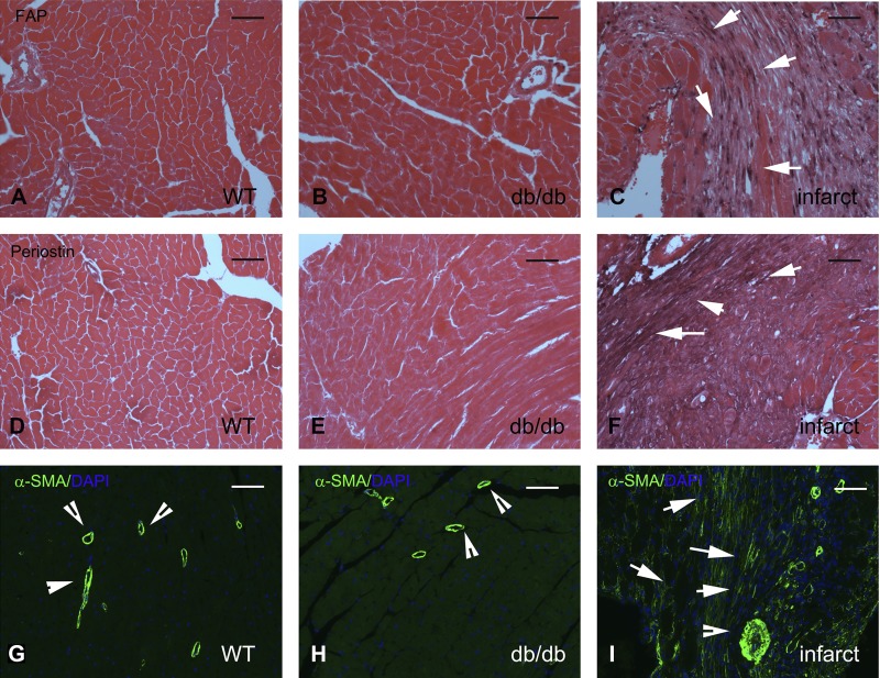
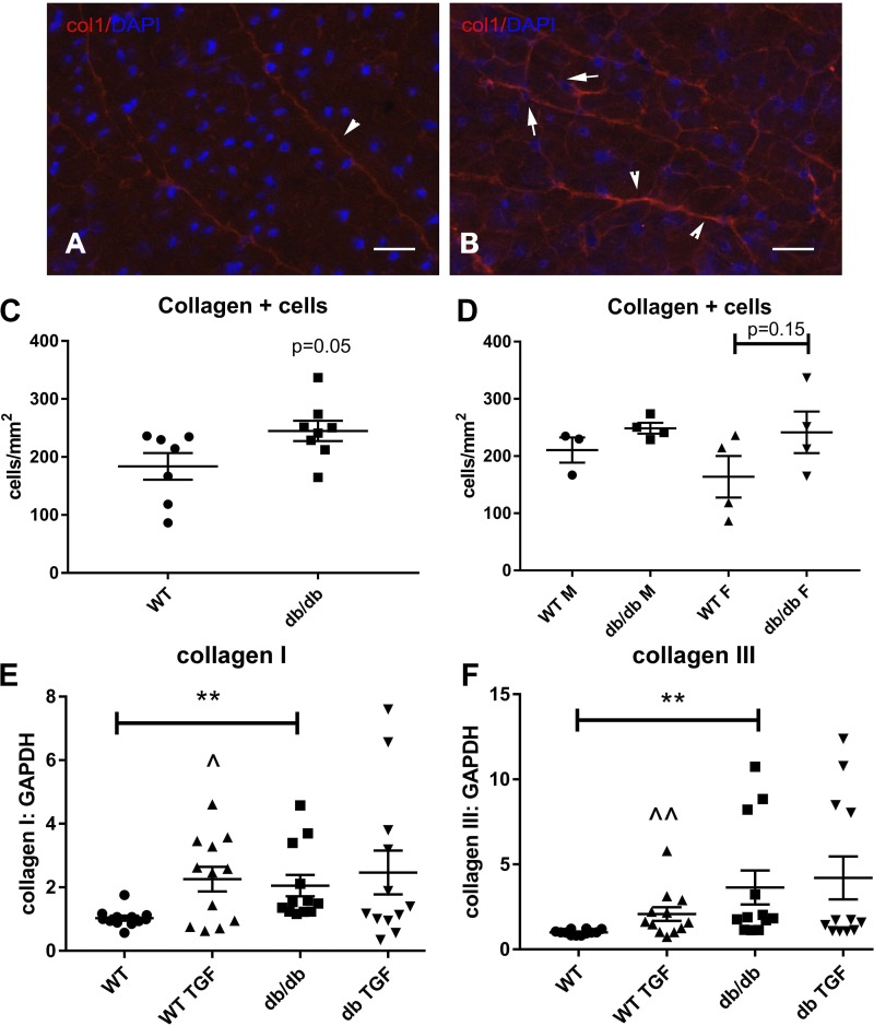
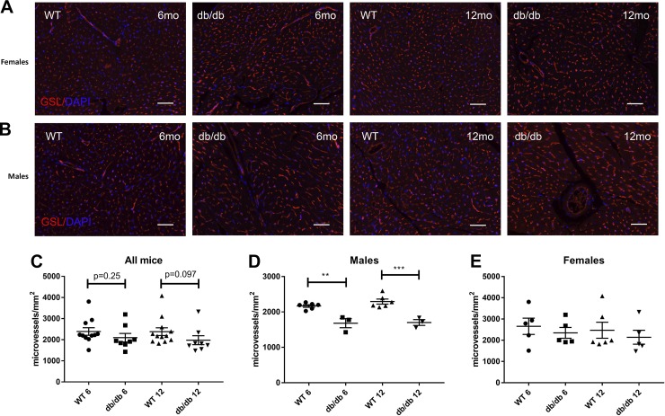
Heart failure with preserved ejection fraction (HFpEF) is caused, or exacerbated by, a wide range of extracardiac conditions. Diabetes, obesity, and metabolic dysfunction are associated with a unique HFpEF phenotype, characterized by inflammation, cardiac fibrosis, and microvascular dysfunction. Development of new therapies for HFpEF is hampered by the absence of reliable animal models. The leptin-resistant db/ db mouse has been extensively studied as a model of diabetes-associated cardiomyopathy; however, data on the functional and morphological alterations in db/ db hearts are conflicting. In the present study, we report a systematic characterization of the cardiac phenotype in db/ db mice, focusing on the time course of functional and histopathological alterations and on the identification of sex-specific cellular events. Although both male and female db/ db mice developed severe obesity, increased adiposity, and hyperglycemia, female mice had more impressive weight gain and exhibited a modest but significant increase in blood pressure. db/ db mice had hypertrophic ventricular remodeling and diastolic dysfunction with preserved ejection fraction; the increase in left ventricular mass was accentuated in female mice. Histological analysis showed that both male and female db/ db mice had cardiomyocyte hypertrophy and interstitial fibrosis, associated with marked thickening of the perimysial collagen, and expansion of the periarteriolar collagen network, in the absence of replacement fibrosis. In vivo and in vitro experiments showed that fibrotic changes in db/ db hearts were associated with increased collagen synthesis by cardiac fibroblasts, in the absence of periostin, α-smooth muscle actin, or fibroblast activation protein overexpression. Male db/ db mice exhibited microvascular rarefaction. In conclusion, the db/ db mouse model recapitulates functional and histological features of human HFpEF associated with metabolic dysfunction. Development of fibrosis in db/ db hearts, in the absence of myofibroblast conversion, suggests that metabolic dysfunction may activate an alternative profibrotic pathway associated with accentuated extracellular matrix protein synthesis. NEW & NOTEWORTHY We provide a systematic analysis of the sex-specific functional and structural myocardial alterations in db/ db mice. Obese diabetic C57BL6J db/ db mice exhibit diastolic dysfunction with preserved ejection fraction, associated with cardiomyocyte hypertrophy, interstitial/perivascular fibrosis, and microvascular rarefaction, thus recapitulating aspects of human obesity-related heart failure with preserved ejection fraction. Myocardial fibrosis in db/ db mice is associated with a matrix-producing fibroblast phenotype, in the absence of myofibroblast conversion, suggesting an alternative mechanism of activation.

Keywords: diabetes; diastolic dysfunction; fibroblast; fibrosis; hypertrophy.


Figures

Fig. 1.
Fig. 1.
Both male and female db/db (db) mice in a C57BL6J background exhibit severe obesity, increased adiposity, and overt diabetes. AC: body weight (BW) was markedly higher in db/db mice compared with age-matched wild-type (WT) control mice. Compared with age-matched WT mice, female db/db mice had a more impressive increase in body weight than male mice (female db/db mice: 2.38-fold higher body weight than age-matched WT mice at 6 mo of age and 2.37-fold higher at 12 mo vs. male db/db mice: 1.88-fold at 6 mo and 1.74-fold at 12 mo of age). DF: magnetic resonance imaging showed a marked increase in body fat weight in both male and female db/db mice at 2 and 6 mo of age. GI: in contrast, lean mass was comparable between age- and sex-matched WT and db/db mice. J and K: dual-energy X-ray absorptiometry performed at 4 mo of age also showed that db/db mice had a marked increase in total (G) and abdominal (H) fat content. LN: although there was significant variability, both male (M) and female (N) db/db animals exhibited a marked increase in fasting plasma glucose levels at 2 and 6 mo of age. **P < 0.01, ***P < 0.001, ****P < 0.0001, and ^P < 0.05 vs. corresponding 2-mo-old mice; ^^^P < 0.001 vs. corresponding 2-mo-old mice. Body weight sample size: WT mice: 1 mo n = 37, 2 mo n = 63, 4 mo n = 58, 6 mo n = 91, 12 mo n = 22; db/db mice: 1 mo n = 11, 2 mo n = 35, 4 mo n = 45, 6 mo n = 68, 12 mo n = 24. Male mice: WT 1 mo n = 16, 2 mo n = 23, 4 mo n = 32, 6 mo n = 53, 12 mo n = 11; db/db: 1 mo n = 7, 2 mo n = 22, 4 mo n = 29, 6 mo n = 38, 12 mo n = 10. Female mice: WT 1 mo n = 21, 2 mo n = 40, 4 mo n = 31, 6 mo n = 38, 12 mo n = 11; db/db: 1 mo n = 4, 2 mo n = 13, 4 mo n = 16, 6 mo n = 30, 12 mo n = 14. Magnetic resonance imaging fat content: n = 12–16 mice/group, n = 5–9 male mice/group, n = 4–8 female mice/group. Dual-energy X-ray absorptiometry: n = 7–16/group. Fasting plasma glucose: n = 18–49 mice/group, n = 9–27 male mice/group, and n = 9–22 female mice/group.
Fig. 2.
Fig. 2.
Female db/db mice have a modest but significant elevation in systolic blood pressure (SBP) and diastolic blood pressure (DBP). A and B: at 4 mo of age, obese diabetic db/db mice had a significant elevation in SBP and a trend toward higher DBP. C and D: sex-specific analysis showed that male db/db mice had a trend toward increased SBP (C) and no significant difference in DBP (D) compared with lean WT control mice. Female db/db mice had significantly increased SBP (C) and DBP (D) compared with age-matched WT control mice (n = 10–11 male mice/group, n = 7–8 female mice/group, n = 18 male + female mice/group). *P < 0.05 and **P < 0.01. BP, blood pressure.
Fig. 3.
Fig. 3.
Obese diabetic db/db mice exhibit cardiac remodeling with preserved ejection fraction. A: echocardiographic imaging was performed in wild-type (WT) and db/db mice. Representative images show evidence of cardiac remodeling in db/db mice at 6 mo of age compared with lean WT control mice (scale bar = 2 mm). B: db/db mice had significantly higher left ventricular (LV) mass at 2 and 6 mo of age. C and D: both male and female db/db mice had increased LV mass at 6 mo of age. However, the increase in LV mass was exaggerated in female db/db mice. At 6 mo of age, male db/db mice had a 15.1% higher LV mass than age-matched WT mice, whereas female db/db animals at the same age exhibited a 46.4% increase. E: LV end-diastolic volume (LVEDV) was increased in db/db mice. F and G: female mice had earlier ventricular dilation than male mice. HJ: obese diabetic db/db mice exhibited slightly but significantly higher ejection fraction at 6 mo of age than age-matched WT control mice. KM: the global remodeling index was assessed to compare maladaptive hypertrophic remodeling between groups. K: at 6 mo of age, db/db mice had a lower remodeling index than corresponding lean WT control mice. L and M: female but not male db/db mice had a significant reduction in the global remodeling index, indicating accentuated hypertrophic remodeling (n = 19–44 male mice/group, n = 12–37 female mice/group, n = 29–81 male + female mice/group). *P < 0.05, **P < 0.01, ***P < 0.001, and ****P < 0.0001.
Fig. 4.
Fig. 4.
Tissue Doppler imaging suggests that db/db mice develop diastolic dysfunction. AC: both male and female db/db mice and age-matched wild-type (WT) control mice had comparable heart rates. DF: mitral inflow Doppler showed no significant differences in the E-to-A ratio between groups. GI: tissue Doppler imaging showed that the e′-to-a′ ratio was significantly reduced in both male and female db/db mice at 6 mo of age. JL: the E-to-e′ ratio was significantly increased in both male and female db/db mice at 6 mo of age. M: representative images of tissue Doppler tracings showing the changes in the e′-to-a′ ratio in 6-mo-old db/db mice compared with age-matched lean control mice. These findings suggest that db/db mice develop diastolic dysfunction. Tissue Doppler imaging may be more sensitive than mitral inflow Doppler in detecting changes in diastolic function in mice (n = 7–19 male mice/group, n = 7–16 female mice/group, n = 14–35 male + female mice/group). *P < 0.05, **P < 0.01, ***P < 0.001, and ****P < 0.0001 vs. corresponding lean mice; ^P < 0.05, ^^P < 0.01, and ^^^P < 0.0001 vs. corresponding 2-mo-old mice.
Fig. 5.
Fig. 5.
Obese diabetic db/db mice had significantly higher left ventricular end-diastolic pressure (LVEDP) than lean wild-type (WT) mice at 6 mo of age. A: db/db mice had significantly higher LVEDP than age-matched lean WT mice. B and C: sex-specific analysis showed that male db/db mice had a trend toward increased LVEDP (B), whereas female mice had significantly higher LVEDP (C; n = 6–7 male mice/group, n = 3–7 female mice/group, n = 10–13 male + female mice/group). *P < 0.05.
Fig. 6.
Fig. 6.
Obese diabetic db/db mice exhibit a marked increase in cardiomyocyte size without a significant increase in the density of interstitial cells. A: wheat germ agglutinin (WGA) lectin histochemistry was used to quantitatively assess cardiomyocyte size. DAPI staining was used for the quantification of interstitial cell density. BD: both male and female db/db mice had a marked increase in cardiomyocyte size at 2 and 6 mo of age. EG: interstitial cell density was comparable between groups (n = 4 male mice/group, n = 4 female mice/group, n = 8 male + female mice/group). Scale bar = 50 μm. *P < 0.05, **P < 0.01, and ***P < 0.001.
Fig. 7.
Fig. 7.
Obese diabetic db/db mice do not have replacement fibrosis but exhibit thickening of the perimysial collagen network and increased endomysial collagen. A and B: picrosirius red staining was used to identify perimysial collagen fibers that form the sheath that groups cardiomyocytes into bundles (long arrows) and endomysial collagen fibers surrounding each individual cardiomyocyte (short arrows). There was no evidence of replacement fibrosis in any of the db/db hearts. CE: semiquantitative analysis showed that db/db mice have accentuated endomysial collagen at 6 mo of age. The increased endomysial collagen score in male or female mice did not reach statistical significance. FH: perimysial collagen thickness was markedly increased in db/db mice at 6 and 12 mo of age. Both female and male mice had increased perimysial collagen thickness at 6 mo of age (n = 4 male mice/group, n = 4 female mice/group, n = 8 male + female mice/group). Scale bar = 50 μm. *P < 0.05, **P < 0.01, ***P < 0.001, ^P < 0.05, and ^^P < 0.01 vs. the corresponding 6-mo group.
Fig. 8.
Fig. 8.
db/db mice exhibit expansion of the periadventitial collagen network in coronary arterioles. A and B: periarteriolar collagen was identified using picrosirius red staining in male and female wild-type (WT) and db/db mouse hearts (white arrows). Please note the increased perimysial thickness (arrowheads) and the accentuated deposition of endomysial collagen (black arrow) in db/db mouse hearts (quantified in Fig. 7). C: quantitative analysis showed that at 12 mo of age, the periadventitial collagen area was higher in arterioles of db/db mice than in the corresponding vessels of WT mice. D and E: both male (D) and female (E) animals exhibited expansion of the periadventitial collagen area (n = 19–45 vessels/group for male mice, n = 19–35 vessels/group for female mice, n = 50–70 vessels/group for male + female mice). Scale bar = 50 μm. *P < 0.05, **P < 0.01, and ***P < 0.001.
Fig. 9.
Fig. 9.
Female db/db mice show significant hypertrophy of the coronary arteriolar media. A: quantitative analysis of the picrosirius red-stained sections (shown in Fig. 8) suggested that db/db mice had a significantly higher mean arteriolar area compared with wild-type (WT) mice at 6 and 12 mo of age. B: male mice had a trend toward increased arteriolar area at 12 mo of age. C: female mice had significantly higher arteriolar area at both 6- and 12-mo time points. D and E: α-smooth muscle actin (α-SMA) immunofluorescence showed hypertrophy of the arteriolar media (arrows) in female db/db mice (n = 19–45 vessels/group for male mice, n = 19–35 vessels/group for female mice, n = 50–70 vessels/group for male + female mice). Scale bar = 50 μm. *P < 0.05, **P < 0.01, and ****P < 0.0001.
Fig. 10.
Fig. 10.
Increased collagen deposition in db/db hearts is not associated with myofibroblast conversion. AC: immunohistochemical staining for fibroblast activation protein (FAP), a marker for activated fibroblasts, in lean wild-type (WT; A), uninjured diabetic (B), and infarcted WT C57BL6J (C) mouse hearts. No FAP+ cells were noted in WT or diabetic myocardium. In contrast, abundant FAP+ fibroblasts infiltrated the infarcted myocardium 7 days after coronary occlusion (arrows in C). Images were counterstained with eosin. DF: periostin staining in lean WT (D), uninjured diabetic (E), and infarcted WT C57BL6J (F) mouse hearts. In injury sites and in fibrotic tissues, activated myofibroblasts typically exhibited periostin expression. E: please note the complete absence of periostin immunoreactivity in db/db hearts. In contrast, infarcted hearts (F) exhibited periostin expression in activated myofibroblasts and in the surrounding extracellular matrix (arrows). GI: α-smooth muscle actin (α-SMA) immunofluorescence was used to identify activated myofibroblasts as spindle-shaped immunoreactive cells located outside the vascular media. In uninjured WT (G) and db/db (H) hearts, α-SMA was exclusively localized in the arteriolar media (arrowheads). I: please note the abundant α-SMA-expressing myofibroblasts in the infarcted myocardium (arrows). Images show sections from 6-mo-old-mice representative of at least 4 different animals/group. Scale bar = 50 μm.
Fig. 11.
Fig. 11.
Fibroblasts in db/db hearts show increased collagen synthesis. A and B: cryosections from 6-mo-old lean wild-type (WT) and db/db mouse hearts were stained with an anticollagen type I antibody. DAPI counterstaining was used to identify collagen type I-expressing interstitial cells. C: db/db hearts had a higher density of collagen type I-expressing interstitial cells that did not reach statistical significance (n = 8 hearts/group, P = 0.05). D: sex-specific analysis showed a trend toward higher density of collagen type I-expressing cells in female db/db hearts (n = 4 hearts/group). E and F: in vitro, cardiac fibroblasts harvested from 4-mo-old db/db mice had a 2.0- to 3.0-fold increase in baseline collagen type I (E) and type III (F) mRNA expression compared with fibroblasts from WT hearts. Activated fibroblasts from db/db hearts were less responsive to transforming growth factor (TGF)-β1 stimulation. TGF-β1 (10 ng/ml) stimulation for 4 h stimulated collagen type I and type III mRNA synthesis in WT cells but did not significantly increase expression of collagens in db/db fibroblasts (n = 7–8/group). Scale bar = 25 μm. **P < 0.01, ^P < 0.05, and ^^P < 0.01 vs. corresponding unstimulated cells. F, female; M, male.
Fig. 12.
Fig. 12.
Male db/db mice had microvascular rarefaction. A and B: microvascular density was assessed in db/db and lean wild-type (WT) mouse hearts using Griffonia simplicifolia (GSL)-I lectin staining. C: db/db mice had a trend toward reduced microvascular density at 6 and 12 mo of age. D and E: although female db/db mice had comparable microvascular density with age-matched WT control mice, male mice exhibited a markedly lower microvascular density (n = 4 male mice/group, n = 4 female mice/group, n = 8 male + female mice/group). Scale bar = 50 μm. **P < 0.01 and ***P < 0.001.

References

    1. Abbate A, Arena R, Abouzaki N, Van Tassell BW, Canada J, Shah K, Biondi-Zoccai G, Voelkel NF. Heart failure with preserved ejection fraction: refocusing on diastole. Int J Cardiol 179: 430–440, 2015. doi:10.1016/j.ijcard.2014.11.106. - DOI - PubMed
    1. Alvarez JA, Reyes M, Escobedo D, Freeman GL, Steinhelper ME, Feldman MD. Enhanced left ventricular systolic function early in type 2 diabetic mice: clinical implications. Diab Vasc Dis Res 1: 89–94, 2004. doi:10.3132/dvdr.2004.013. - DOI - PubMed
    1. Barouch LA, Gao D, Chen L, Miller KL, Xu W, Phan AC, Kittleson MM, Minhas KM, Berkowitz DE, Wei C, Hare JM. Cardiac myocyte apoptosis is associated with increased DNA damage and decreased survival in murine models of obesity. Circ Res 98: 119–124, 2005. doi:10.1161/01.RES.0000199348.10580.1d. - DOI - PubMed
    1. Biernacka A, Cavalera M, Wang J, Russo I, Shinde A, Kong P, Gonzalez-Quesada C, Rai V, Dobaczewski M, Lee DW, Wang XF, Frangogiannis NG. Smad3 signaling promotes fibrosis while preserving cardiac and aortic geometry in obese diabetic mice. Circ Heart Fail 8: 788–798, 2015. doi:10.1161/CIRCHEARTFAILURE.114.001963. - DOI - PMC - PubMed
    1. Buckley LF, Canada JM, Del Buono MG, Carbone S, Trankle CR, Billingsley H, Kadariya D, Arena R, Van Tassell BW, Abbate A. Low NT-proBNP levels in overweight and obese patients do not rule out a diagnosis of heart failure with preserved ejection fraction. ESC Heart Fail 5: 372–378, 2018. doi:10.1002/ehf2.12235. - DOI - PMC - PubMed

Publication types

MeSH terms

Substances

LinkOut - more resources