. 2018 Aug 7;57(31):4675-4689.
doi: 10.1021/acs.biochem.7b01081. Epub 2018 Jul 26.

An Open Library of Human Kinase Domain Constructs for Automated Bacterial Expression

Affiliations

An Open Library of Human Kinase Domain Constructs for Automated Bacterial Expression

Steven K Albanese et al. Biochemistry. .

Abstract

Kinases play a critical role in cellular signaling and are dysregulated in a number of diseases, such as cancer, diabetes, and neurodegeneration. Therapeutics targeting kinases currently account for roughly 50% of cancer drug discovery efforts. The ability to explore human kinase biochemistry and biophysics in the laboratory is essential to designing selective inhibitors and studying drug resistance. Bacterial expression systems are superior to insect or mammalian cells in terms of simplicity and cost effectiveness but have historically struggled with human kinase expression. Following the discovery that phosphatase coexpression produced high yields of Src and Abl kinase domains in bacteria, we have generated a library of 52 His-tagged human kinase domain constructs that express above 2 μg/mL of culture in an automated bacterial expression system utilizing phosphatase coexpression (YopH for Tyr kinases and lambda for Ser/Thr kinases). Here, we report a structural bioinformatics approach to identifying kinase domain constructs previously expressed in bacteria and likely to express well in our protocol, experiments demonstrating our simple construct selection strategy selects constructs with good expression yields in a test of 84 potential kinase domain boundaries for Abl, and yields from a high-throughput expression screen of 96 human kinase constructs. Using a fluorescence-based thermostability assay and a fluorescent ATP-competitive inhibitor, we show that the highest-expressing kinases are folded and have well-formed ATP binding sites. We also demonstrate that these constructs can enable characterization of clinical mutations by expressing a panel of 48 Src and 46 Abl mutations. The wild-type kinase construct library is available publicly via Addgene.


Figures

Figure 1
Figure 1. Abl kinase domain construct expression screen illustrates high sensitivity to construct boundaries
(A) Abl kinase domain construct boundaries with highest expression yields. Standard deviations of the yield are listed for control constructs for which six replicates were performed to give an indication of the uncertainty in experimental constructs. Secondary structure is indicated on the sequence. Beta sheets are colored blue and alpha helices are colored orange. (B) Heatmap showing average yields for constructs (in μg/mL culture) with detectable expression as a function of N- and C-terminal construct boundaries. (C) left: PDBID: 2E2B with the nine N-terminal construct boundary amino acids shown as yellow spheres. right: PDBID: 4XEY with the nine C-terminal construct boundary amino acids shown as green spheres. Black arrows indicate residue numbers.
Figure 2
Figure 2. Expression yields of Abl kinase domain constructs for all constructs with detectable expression
A synthetic gel image rendering generated from Caliper GX II microfluidic gel electrophoresis data following Ni-affinity purification and thermal denaturation for all Abl constructs with detectable expression. Each well is marked with the Abl kinase domain construct residue boundaries (Uniprot canonical isoform numbering). Bands for YopH164 phosphatase (50 kDA) and Abl kinsase domain constructs (28–35 kDA) are labeled.
Figure 3
Figure 3. Kinome wide search for expressible kinases
(A) The number of PDB structures per kinase family, from the database built to select kinases for expression. (B) The distribution among familes of candidate kinases in our expression screen. (C) Caliper GX II synthetic gel image rendering of the highest expressing kinases, quantified using microfluidic capillary electrophoresis. (D) Kinome distribution of expression based on our 96 kinase screen. Dark green circles represent kinases with expression above 50 μg/mL culture yield. Light green circles represent kinases with expression between 50 and 12 μg/mL yield. Yellow circles represent kinases with expression between 12 and 7 μg/mL yield. Orange circles represent kinases with any expression (even below 2 μg/mL) up to 7 μg/mL yield. Image made with KinMap: http://www.kinhub.org/kinmap.
Figure 4
Figure 4. Fluorescence-based thermostability assay demonstrates many high-expressing kinases are well-folded
A fluorescence-based thermostability assay was performed on the 14 kinases shown to express above a minimum 0.24 mg/mL concentration after elution. SYPRO Orange fluorescence (solid blue line) was measured at 580 nm (half bandwidth 20 nm) after excitation at 465 nm (half bandwith 25 nm) as as the temperature was ramped from (x-axis) in Nickel Buffer A (25 mM HEPES pH 7.5, 5% glycerol, 400 mM NaCl, 20 mM imidazole, 1 mM BME). The temperature was held at 25°C for 15 sec before ramping up to 95°C with a ramp rate of 0.06°C/s. The unfolding temperature Tm (black dashed line and insert) was determined from the maxima of the normalized first derivative of fluorescence (red dashed line). Fluorescence emission at 580 nm is shown on the left y-axis. To control for signals resulting from TEV protease contamination present at 0.01–0.03 mg/mL, TTK, a kinase with no detectable expression in our panel as determined via Caliper GX II quantitation was in included (panel 15).
Figure 5
Figure 5. Fluorescence emission spectra as a function of the fluorescent ATP-competitive kinase inhibitor bo-sutinib demonstrates the presence of a well-formed ATP binding pocket
The ATP-competitive inhibitor bosutinib shows a strong increase in fluorescence centered around 450 nm when bound to kinases with well-folded ATP binding sites upon excitation at 280 nm. To assess whether the kinases from the high-throughput expression screen were well-folded, bosutinib was titrated in a 15-concentration series geometrically spanning 0.008 μM to 18.99 μM (colored lines, higher concentrations are shown in warmer colors) in 15 increments for 39 expressing kinases with protein concentration adjusted to ~0.5 μM in 100 μL assay volume. Eluted TEV protease contaminant varies from 0.01–0.03 mg/mL in the assay volumes. The control MK14 and boiled MK14 (boiled for 10 min at 95°C) were produced in a large scale expression from the same plasmid as used in the high-throughput expression protocol and they were included as positive and negative controls for bosutinib binding to ATP binding pocket. Fluorescence emission spectra (y-axis, bandwidth 20 nm) were measured from 370 nm to 600 nm (x-axis) for excitation at 280 nm (bandwidth 10 nm). For reference, the fluorescence of bosutinib titrated into buffer titration (panel 36) is shown in grayscale in each panel. Significant increases in fluorescence signal above baseline qualitatively indicate the presence of a well-formed ATP binding site.
Figure 6
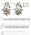
Figure 6. Expression yields for engineered clinically-derived Src and Abl missense mutants
(A) All Abl and Src clinically-identified mutants assessed in the expression screen are displayed as sticks. Mutants with expression yields >2 μg/mL are colored green, while those with yields <2 μg/mL are colored orange. Rendered structures are Abl (PDBID: 2E2B) and Src (PDBID: 4MXO). (B) Synthetic gel images showing ABl (top) or Src (bottom) expression, with wells labeled by missense mutation. Yield was determined by Caliper GX II quantitation of the expected size band and reported in μg/mL culture, where total eluate volume was 120 μL following nickel bead pulldown purification from 900 μL bacterial culture. Residue mutations use numbering for the Uniprot canonical isoform.

References

    1. Manning G, Whyte DB, Martinez R, Hunter T, Sudarsanam S. Science. 2002;298:1912–1934. - PubMed
    1. Hunter T, Manning G. In: Receptor Tyrosine Kinases: Structure, Functions and Role in Human Disease. Wheeler DL, Yarden Y, editors. Springer; New York: New York, NY: 2015. pp. 1–15.
    1. Knight ZA, Lin H, Shokat KM. Nature Reviews Cancer. 2010;10:130–137. - PMC - PubMed
    1. American Cancer Society. Cancer Facts & Figures 2015. 2015.
    1. Cohen P, Tcherpakov M. Cell. 2010;143:686–693. - PubMed

Publication types

Substances