. 2018 Nov;36(11):1676-1684.
doi: 10.1002/stem.2888. Epub 2018 Jul 29.

JAK2V617F Megakaryocytes Promote Hematopoietic Stem/Progenitor Cell Expansion in Mice Through Thrombopoietin/MPL Signaling

Affiliations

JAK2V617F Megakaryocytes Promote Hematopoietic Stem/Progenitor Cell Expansion in Mice Through Thrombopoietin/MPL Signaling

Yu Zhang et al. Stem Cells. 2018 Nov.

Abstract

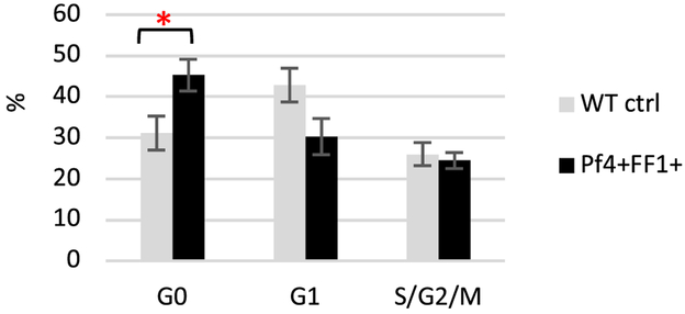
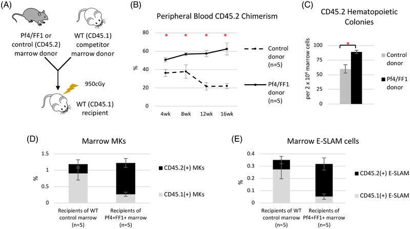
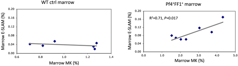
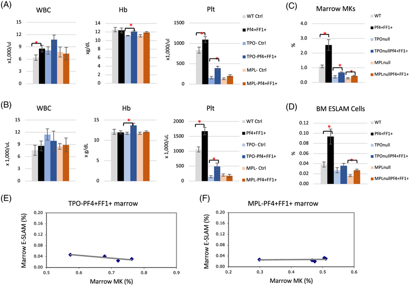
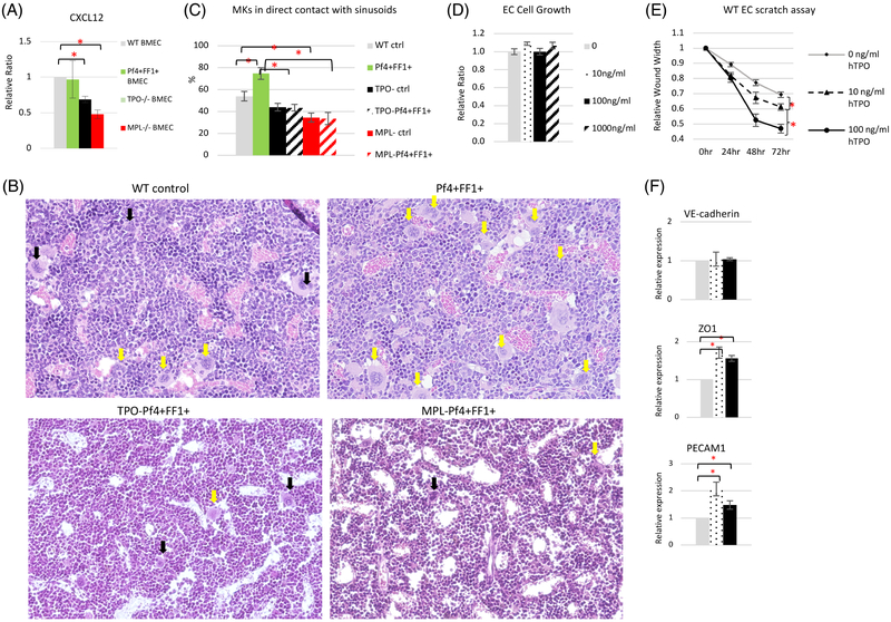
The myeloproliferative neoplasms (MPNs) are stem cell disorders characterized by hematopoietic stem/progenitor cell (HSPC) expansion and overproduction of mature blood cells. The acquired kinase mutation JAK2V617F plays a central role in these disorders. The mechanisms responsible for HSPC expansion in MPNs are not fully understood, limiting the effectiveness of current treatments. One hallmark feature of the marrow in patients with MPNs is megakaryocyte (MK) hyperplasia. Previously, we reported that JAK2V617F-bearing MKs cause a murine myeloproliferative syndrome with HSPC expansion. Here we show that JAK2V617F MKs promote MPN stem cell function by inducing HSPC quiescence with increased repopulating capacity. In addition, we demonstrate that thrombopoietin and its receptor MPL are critical for the JAK2V617F-bearing MK-induced myeloproliferation, both by directly affecting the quantity and quality of MKs and by altering the MK-endothelial interaction and vascular niche function. Therefore, targeting HSPC niche-forming MKs and/or their interactions within the vascular niche could provide novel, more effective therapeutic strategies in patients with MPNs. Stem Cells 2018;36:1676-1684.

Keywords: JAK2; MPL; Megakaryocyte; Myeloproliferative neoplasm; Thrombopoietin.


Conflict of interest statement

CONFLICT OF INTERST

The authors declare no conflict of interest.

Figures

Figure 1.
Figure 1.
HSPCs in the JAK2V617F-bearing MK niche are more quiescent than in the WT MK niche. Cell cycle analysis of marrow CD150+CD48 HSPCs from WT control mice and Pf4+FF1+ mice. Data are presented as mean ±SEM; n = 4 mice per group. *P < 0.05.
Figure 2.
Figure 2.
The JAK2V617F-bearing MK niche promotes the expansion/engraftment of JAK2V617F HSPCs. (A) Competitive marrow transplantation scheme. (B) Following the competitive transplantation experiment, recipients of the Pf4+FF1+ marrow (n = 5) displayed a higher peripheral blood donor (CD45.2) chimerism than the recipients of the control marrow (n = 5). (C) There was a moderate increase of CD45.2+ hematopoietic progenitors in Pf4+FF1+ marrow recipients compared to control marrow recipients. (D) CD45.2+ MKs were significantly increased while CD45.1+ MKs were significantly decreased in the Pf4+FF1+ marrow recipients compared to controls. (E) CD45.2+ E-SLAM cells were significantly increased while CD45.1+ E-SLAM cells were significantly decreased in recipients of Pf4+FF1+ marrow compared to controls. Data are presented as mean ±SEM. *P < 0.05.
Figure 3.
Figure 3.
Marrow E-SLAM cell numbers correlate strongly with marrow MK cell numbers in the Pf4+FF1+ mice, but not in the WT control mice. Marrow MK and E-SLAM cell frequencies were measured by flow cytometry analysis. Left: WT control mice (n = 6); Right: Pf4+FF1+ mice (n = 7).
Figure 4.
Figure 4.
TPO/MPL signaling is required for the development of JAK2V617F MK-induced myeloproliferation. (A-B) Peripheral blood cell counts in WT control (Pf4+FF1), Pf4+FF1+, TPO control (TPOPf4+FF1), TPOPf4+FF1+, MPL control (MPLPf4+FF1), and MPLPf4+FF1+ mice at 28 weeks of age (A) and 1 year of age (B). Data are presented as mean ±SEM; n = 6–11 mice per group. (C-D) At 28 weeks of age, marrow MKs (C) and marrow E-SLAM cells (D) were significantly increased in the Pf4+FF1+ mice compared to WT controls, but not in the TPOPf4+FF1+ mice or MPLPf4+FF1+ mice. Data are presented as mean ±SEM; n = 4–8 mice per group. (E-F) There was no correlation between Marrow MK and E-SLAM cell frequencies in TPOPf4+FF1+ (n = 4) (E) or MPLPf4+FF1+ (n = 5) mice (F). Data are presented as mean ±SEM. *P < 0.05.
Figure 5.
Figure 5.
TPO and MPL are important for vascular niche function. (A) The expression levels of CXCL12 in marrow ECs isolated from WT ctrl, Pf4+FF1+, TPO−/−, and MPL−/− mice. Gene expression is shown as the relative fold-change compared to WT ctrl marrow EC expression which was set as ‘1’. (B) Representative images of H&E sections of femoral marrow of Pf4-cre WT ctrl mice, Pf4+FF1+ mice, TPOPf4+FF1+ mice and MPLPf4+FF1+ mice. Yellow arrows indicate examples of MKs physically associated with marrow sinusoids. Black arrows indicate examples of MKs not in direct contact with marrow sinusoids. Magnification: x20 (C) Quantification of MKs physically associated with marrow sinusoids in WT ctrl mice (n = 3), Pf4+FF1+ mice (n = 3), TPO ctrl mice (n = 2), TPOPf4+FF1+ mice (n = 2), MPL ctrl mice (n = 2), and MPLPf4+FF1+ mice (n = 2). About 100 marrow MKs from 9–18 non-overlapping fields were studied. (D) TPO did not affect primary murine spleen EC cell growth in vitro. (E) TPO stimulated primary murine spleen EC cell migration in vitro. A lesion was produced across the primary murine spleen EC monolayer in the presence of different TPO concentrations. The distances from one side of the scratch wound to the other side were measured using ImageJ software (National Institute of Health, Bethesda, MD, USA) at six different locations for each culture condition. The distance of wound closure at time 24, 48, and 72 h was compared with the distance at time 0 h which was set as 1. The results were expressed as the mean ±s.e.m. (n = 6). Data are from one of two independent experiments that gave similar results. (F) The expression levels of VE-cadherin, ZO1, and PECAM1 in WT murine lung EC treated with different TPO concentrations. Data are presented as mean ±SEM. *P < 0.05.

References

    1. Walkley CR, Olsen GH, Dworkin S et al. A microenvironment-induced myeloproliferative syndrome caused by retinoic acid receptor gamma deficiency. Cell 2007;129(6):1097–1110. 10.1016/j.cell.2007.05.014. - DOI - PMC - PubMed
    1. Schepers K, Pietras EM, Reynaud D et al. Myeloproliferative neoplasia remodels the endosteal bone marrow niche into a self-reinforcing leukemic niche. Cell Stem Cell 2013;13(3):285–299. 10.1016/j.stem.2013.06.009. - DOI - PMC - PubMed
    1. Arranz L, Sanchez-Aguilera A, Martin-Perez D et al. Neuropathy of haematopoietic stem cell niche is essential for myeloproliferative neoplasms. Nature 2014;512(7512):78–81. 10.1038/nature13383. - DOI - PubMed
    1. Mager LF, Riether C, Schurch CM et al. IL-33 signaling contributes to the pathogenesis of myeloproliferative neoplasms. The Journal of clinical investigation 2015;125(7):2579–2591. 10.1172/JCI77347. - DOI - PMC - PubMed
    1. Walkley CR, Shea JM, Sims NA et al. Rb regulates interactions between hematopoietic stem cells and their bone marrow microenvironment. Cell 2007;129(6):1081–1095. 10.1016/j.cell.2007.03.055. - DOI - PMC - PubMed

Publication types

MeSH terms