. 2018 Oct;11(5):1065-1073.
doi: 10.1016/j.tranon.2018.06.009. Epub 2018 Jul 9.

Molecular Imaging of Pancreatic Duct Adenocarcinoma Using a Type 2 Cannabinoid Receptor-Targeted Near-Infrared Fluorescent Probe

Affiliations

Molecular Imaging of Pancreatic Duct Adenocarcinoma Using a Type 2 Cannabinoid Receptor-Targeted Near-Infrared Fluorescent Probe

Xiaoxia Guo et al. Transl Oncol. 2018 Oct.

Abstract

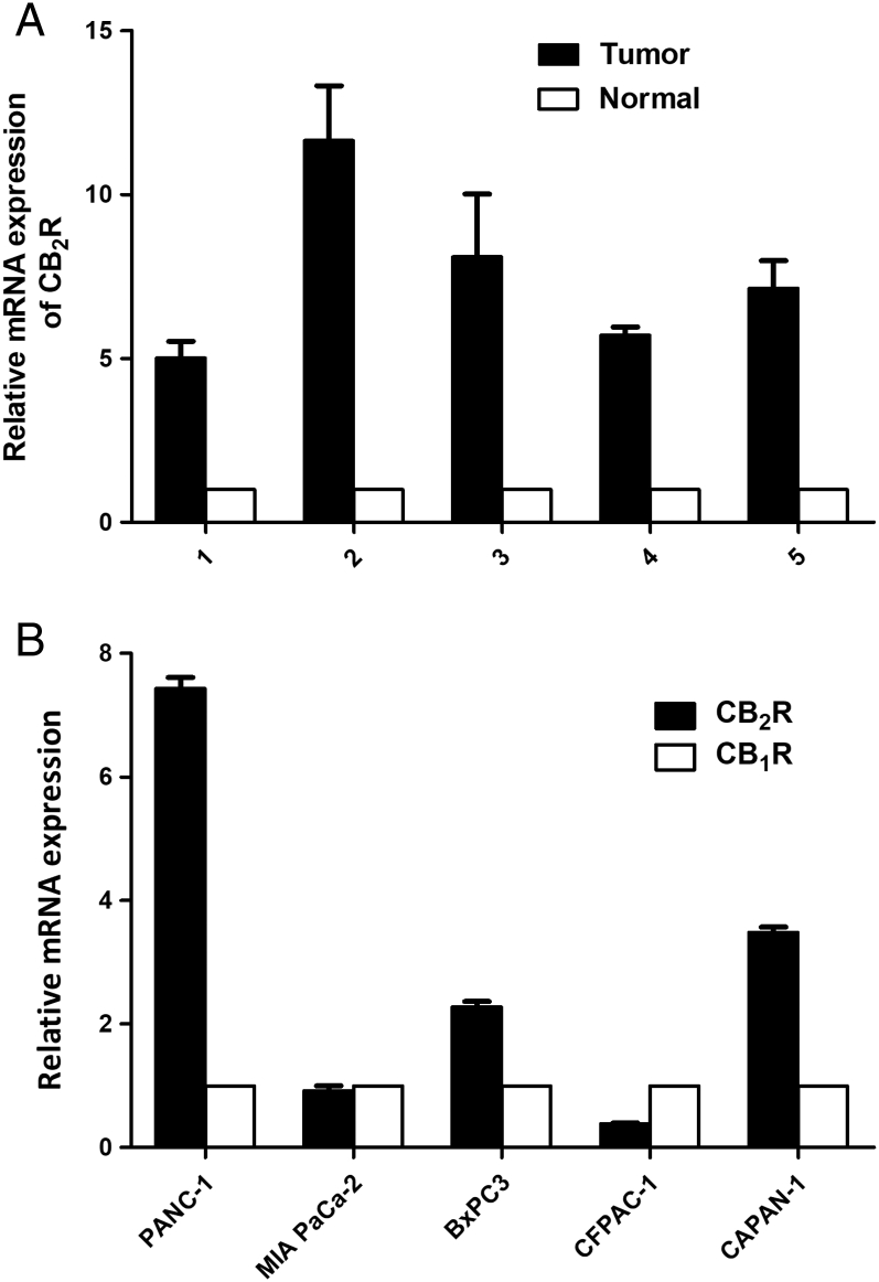
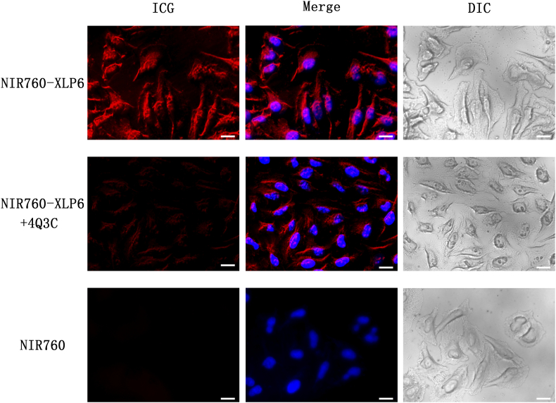
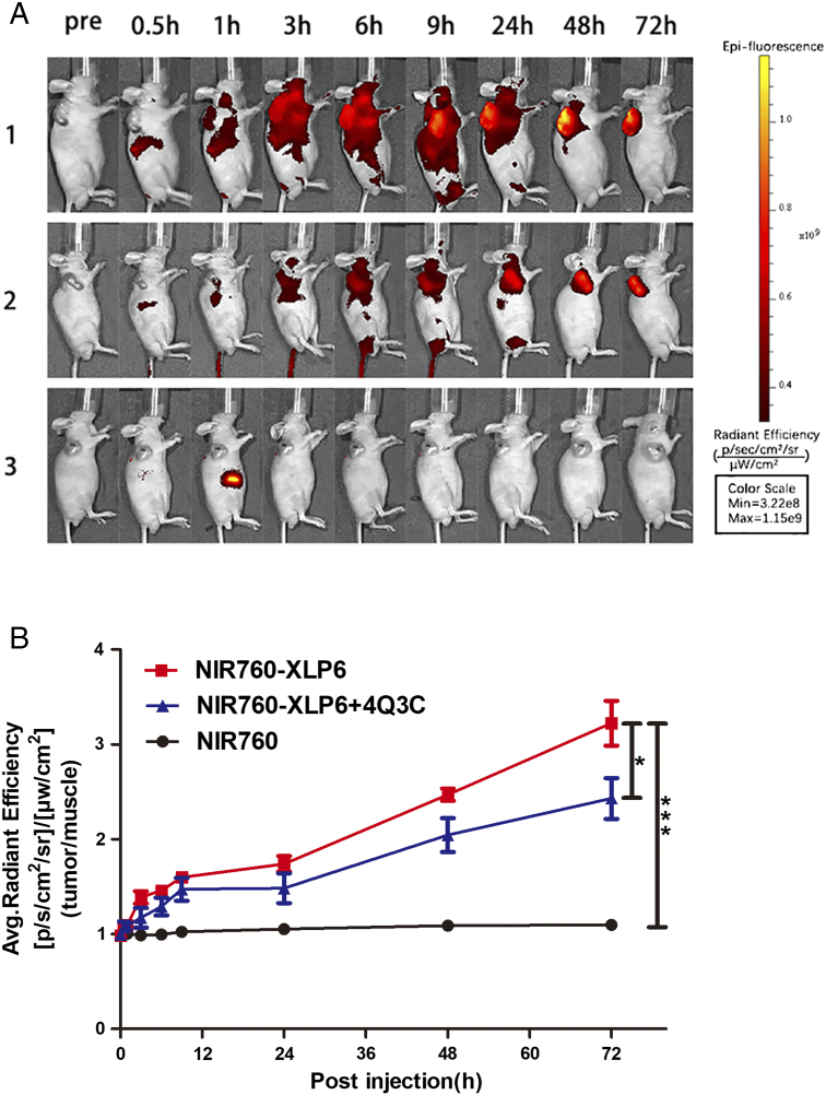
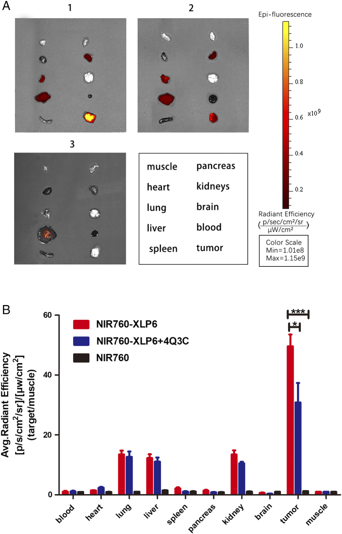
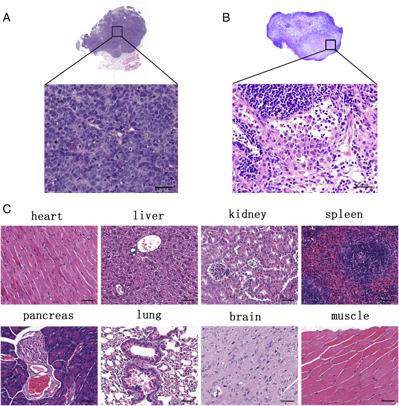
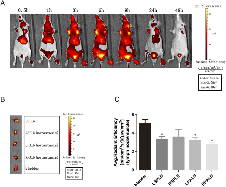
Imaging probes targeting type 2 cannabinoid receptor (CB2R) overexpressed in pancreatic duct adenocarcinoma (PDAC) tissue have the potential to improve early detection and surgical outcome of PDAC. The aim of our study was to evaluate the molecular imaging potential of a CB2R-targeted near-infrared (NIR) fluorescent probe (NIR760-XLP6) for PDAC. CB2R overexpression was observed in both PDAC patient tissues and various pancreatic cancer cell lines. In vitro fluorescence imaging indicated specific binding of NIR760-XLP6 to CB2R in human PDAC PANC-1 cells. In a xenograft mouse tumor model, NIR760-XLP6 showed remarkable 50- (ex vivo) and 3.2-fold (in vivo) tumor to normal contrast enhancement with minimal liver and kidney uptake. In a PDAC lymph node metastasis model, significant signal contrast was observed in bilateral axillary lymph nodes with PDAC metastasis after injection of the probe. In conclusion, NIR760-XLP6 exhibits promising characteristics for imaging PDAC, and CB2R appears to be an attractive target for PDAC imaging.


Figures

Figure 1
Figure 1
Expression of CB2R in human PDAC tissues and cell lines. (A) CB2R expression at the mRNA level was assessed in human PDAC tissues and normal pancreatic tissue samples by real-time PCR. (B) CB1R and CB2R expression at the mRNA level was assessed in PDAC cell lines (CAPAN-1, MIA PaCa-2, BxPC3, PANC-1, CFPAC-1) by real-time PCR.
Figure 2
Figure 2
NIR760-XLP6 specifically binds to CB2R in PANC-1 cells. Cells were divided into three groups as follow: (1) PANC-1 cells treated with NIR760-XLP6; (2) PANC-1 cells treated with 4Q3C followed by NIR760-XLP6; (3) PANC-1 cells treated with NIR760. Fluorescent imaging was obtained using Zeiss Axio Observer fluorescent microscope equipped with the ApoTome 2 imaging system. From left to right, ICG filter (red), ICG filter (red) + DAPI filter (blue) merged, DIC. Scale bars = 20 μm.
Figure 3
Figure 3
In vivo optical imaging of NIR760-XLP6 in xenograft tumor model. All mice were divided into three groups as follows: (1) five PANC-1 tumor-bearing mice injected with NIR760-XLP6; (2) five PANC-1 tumor-bearing mice injected with 4Q3C followed by NIR760-XLP6; (3) five PANC-1 tumor-bearing mice injected with NIR760. (A) Mice were anesthetized and imaged with Xenogen IVIS Spectrum imaging system at preinjection and at 0.5, 1, 3, 6, 9, 24, 48, and 72 hours postinjection. (B) Time activity curves of tumor/normal ratio among three groups. The radiant efficiency of the tumor area at the right flank of the animal (T) and of the area at the left flank normal tissue (N) was calculated by the ROI function in the Living Image software. Dividing T by N yielded the contrast between the tumor tissue and the normal tissue (*P < .05, *** P < .001).
Figure 4
Figure 4
Ex vivo tumor optical imaging and biodistribution study of NIR760-XLP6. All mice were divided into three groups as follows: (1) five PANC-1 tumor-bearing mice injected with NIR760-XLP6; (2) five PANC-1 tumor-bearing mice injected with 4Q3C followed by NIR760-XLP6; (3) five PANC-1 tumor-bearing mice injected with NIR760. (A) Ex vivo imaging of excised tumor and organs at 72 hours postinjection from PANC-1 tumor-bearing mice. (B) Graphical quantification of target/normal signal ratios among the three groups (*P < .05, *** P < .001).
Figure 5
Figure 5
Representative histological images of excised tumor, the major organs, and lymph node from mice postinjected with NIR760-XLP6. (A) Tumor H&E staining image. (B) Metastatic lymph node H&E staining image. (C) The major organs’ H&E staining images. Scale bars = 50 μm.
Figure 6
Figure 6
In vivo and ex vivo optical imaging of NIR760-XLP6 in PDAC lymph node metastasis model. (A) Three PDAC lymph node metastasis mice were injected with NIR760-XLP6 and imaged with Xenogen IVIS Spectrum imaging system at 0.5, 1, 3, 6, 9, 24, and 48 hours postinjection. (B) Ex vivo imaging of lymph nodes at 48 hours postinjection from PDAC lymph node metastasis mice. (C) Graphical quantification of target/normal signal ratios among lymph nodes (*P < .05).

References

    1. Siegel RL, Miller KD, Jemal A. Cancer statistics, 2017. CA Cancer J Clin. 2017;67:7–30. - PubMed
    1. Kamisawa T, Wood LD, Itoi T, Takaori K. Pancreatic cancer. Lancet. 2016;388:73–85. - PubMed
    1. Gillen S, Schuster T, Meyer Zum Buschenfelde C, Friess H, Kleeff J. Preoperative/neoadjuvant therapy in pancreatic cancer: a systematic review and meta-analysis of response and resection percentages. PLoS Med. 2010;7 - PMC - PubMed
    1. Konstantinidis IT, Warshaw AL, Allen JN, Blaszkowsky LS, Castillo CF, Deshpande V, Hong TS, Kwak EL, Lauwers GY, Ryan DP. Pancreatic ductal adenocarcinoma: is there a survival difference for R1 resections versus locally advanced unresectable tumors? What is a "true" R0 resection? Ann Surg. 2013;257:731–736. - PubMed
    1. Frampton AE, Gall TM, Krell J, Ahmad R, Jiao LR. Is there a 'margin' for error in pancreatic cancer surgery? Future Oncol. 2013;9:31–34. - PubMed