

Laparoscopic repair with cone-shaped mesh implantation for perineal hernia occurred after laparoscopic abdominoperineal resection
- PMID: 30005361
- PMCID: PMC6036939
- DOI: 10.1016/j.ijscr.2018.06.032
Laparoscopic repair with cone-shaped mesh implantation for perineal hernia occurred after laparoscopic abdominoperineal resection
Abstract
Introduction: Perineal hernia after abdominoperineal resection (APR) is a rare complication, and no standard surgical procedures are established. We describe a simple laparoscopic mesh implantation technique utilizing a large synthetic flat mesh.
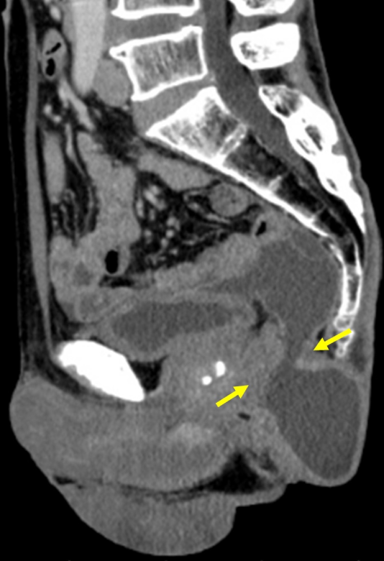
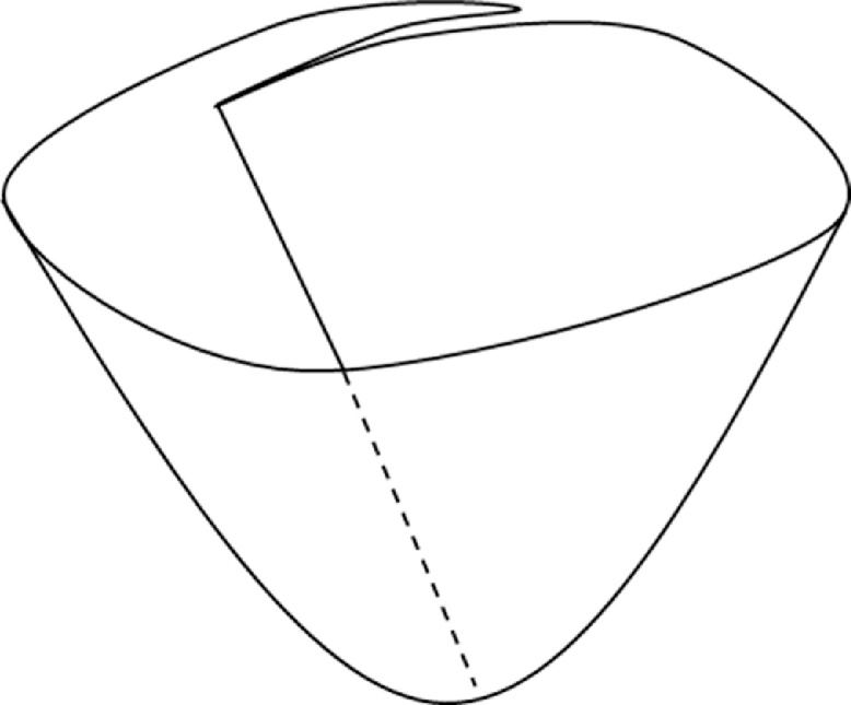
Presentation of case: We report a case of perineal hernia after APR. We performed laparoscopic repair using a soft and large synthetic mesh with simple technique. The essence of this technique is that mesh is inserted into the abdominal cavity without trimming and it forms in a conical shape to better adjust to the pelvic cavity.
Discussion: The perineal and laparoscopic approaches for perineal hernia repair have been performed most commonly in recent years, but the recurrence rate after repair remains high (24.1%). Using a large mesh could cover the hernial orifice with a sufficient margin, reducing a risk of recurrence caused by shrinkage and slippage of the mesh.
Conclusion: Our technique utilizing a large, lightweight, synthetic mesh can be practical and useful for perineal hernia repair after laparoscopic APR.
Keywords: Abdominoperineal resection; Laparoscopy; Mesh repair; Perineal hernia.
Copyright © 2018 The Author(s). Published by Elsevier Ltd.. All rights reserved.
Figures


References
-
- Musters G.D., Buskens C.J., Bemelman W.A., Tanis P.J. Perineal wound healing after abdominoperineal resection for rectal cancer: a systematic review and meta-analysis. Dis. Colon. Rectum. 2014;57:1129–1139. - PubMed
-
- Balla A., Batista Rodriguez G., Buonomo N., Martinez C., Hernandez P., Bollo J. Perineal hernia repair after abdominoperineal excision or extralevator abdominoperineal excision: a systematic review of the literature. Tech. Coloproctol. 2017;21:329–336. - PubMed
-
- Agha R.A., Fowler A.J., Saeta A., Barai I., Rajmohan S., Orgill D.P. The SCARE statement: consensus-based surgical case report guidelines. Int. J. Surg. 2016;34:180–186. - PubMed
-
- Casasanta M., Moore L.J. Laparoscopic repair of a perineal hernia. Hernia. 2012;16:363–367. - PubMed
-
- Dulucq J.L., Wintringer P., Mahajna A. Laparoscopic repair of postoperative perineal hernia. Surg. Endosc. 2006;20:414–418. - PubMed
LinkOut - more resources
Full Text Sources
Other Literature Sources
Research Materials
Miscellaneous
