. 2018 Jul 13;19(1):179.
doi: 10.1186/s12882-018-0968-4.

Angiotensin II-induced podocyte apoptosis is mediated by endoplasmic reticulum stress/PKC-δ/p38 MAPK pathway activation and trough increased Na+/H+ exchanger isoform 1 activity

Affiliations

Angiotensin II-induced podocyte apoptosis is mediated by endoplasmic reticulum stress/PKC-δ/p38 MAPK pathway activation and trough increased Na+/H+ exchanger isoform 1 activity

Vanessa Gerolde Cardoso et al. BMC Nephrol. .

Abstract

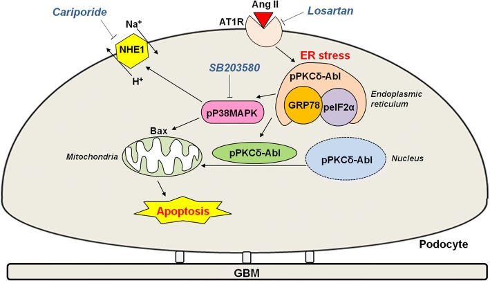
Background: Angiotensin II (Ang II) contributes to the progression of renal diseases associated with proteinuria and glomerulosclerosis mainly by inducing podocyte apoptosis. In the present study, we investigated whether the chronic effects of Ang II via AT1 receptor (AT1R) would result in endoplasmic reticulum (ER) stress/PKC-delta/p38 MAPK stimulation, and consequently podocyte apoptosis.

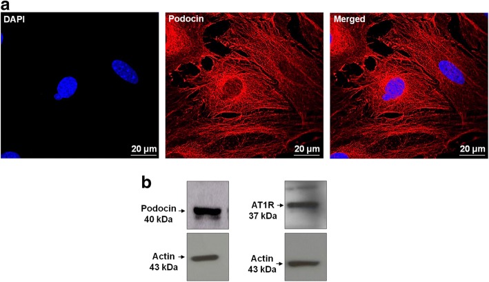
Methods: Wistar rats were treated with Ang II (200 ng·kg-1·min-1, 42 days) and or losartan (10 mg·kg-1·day-1, 14 days). Immortalized mouse podocyte were treated with 1 μM Ang II and/or losartan (1 μM) or SB203580 (0.1 μM) (AT1 receptor antagonist and p38 MAPK inhibitor) for 24 h. Kidney sections and cultured podocytes were used to evaluate protein expression by immunofluorescence and immunoblotting. Apoptosis was evaluated by flow cytometry and intracellular pH (pHi) was analyzed using microscopy combined with the fluorescent probe BCECF/AM.

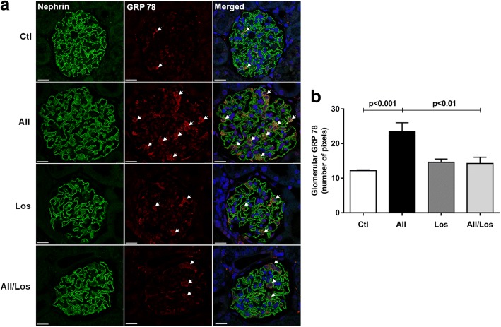
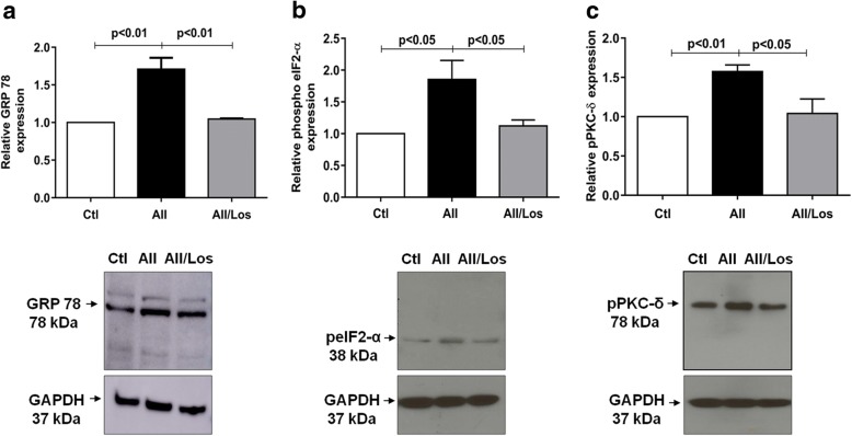
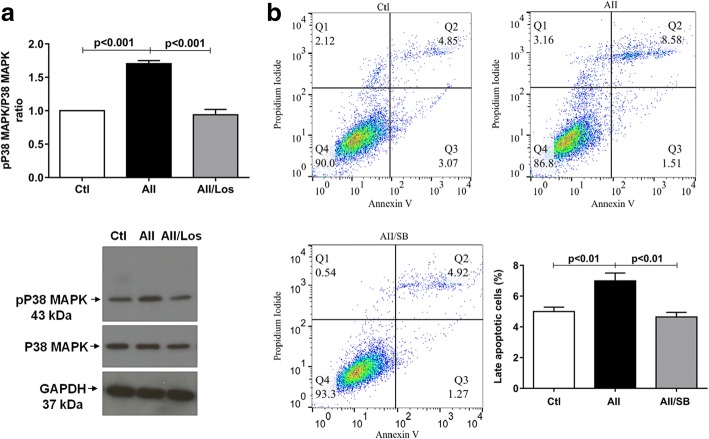
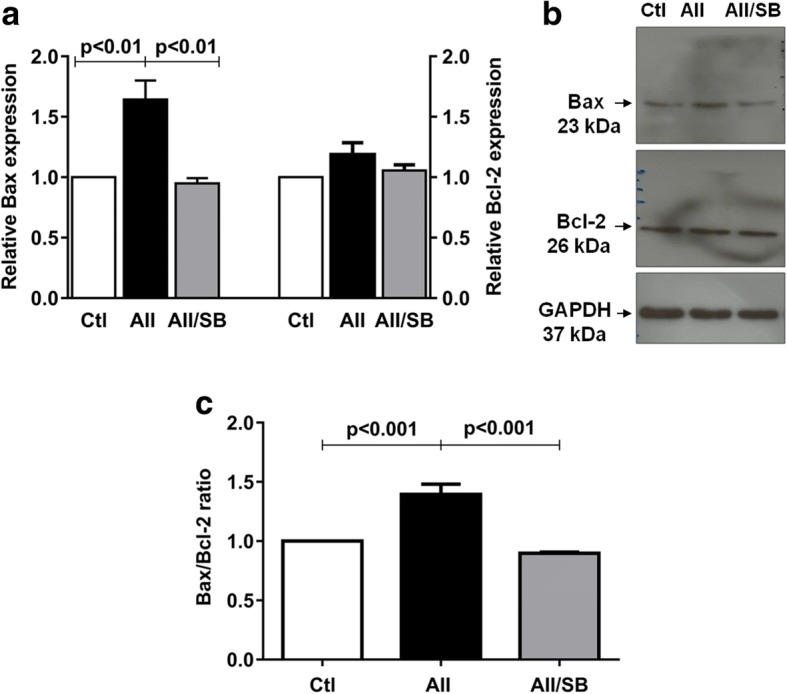
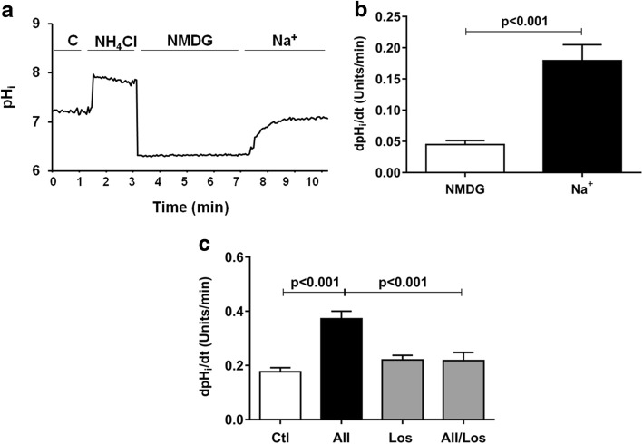
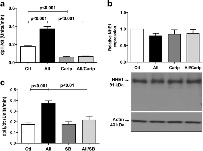
Results: Compared with controls, Ang II via AT1R increased chaperone GRP 78/Bip protein expression in rat glomeruli (p < 0.001) as well as in podocyte culture (p < 0.01); increased phosphorylated eIf2-α (p < 0.05), PKC-delta (p < 0.01) and p38 MAPK (p < 0.001) protein expression. Furthermore, Ang II induced p38 MAPK-mediated late apoptosis and increased the Bax/Bcl-2 ratio (p < 0.001). Simultaneously, Ang II via AT1R induced p38 MAPK-NHE1-mediated increase of pHi recovery rate after acid loading.

Conclusion: Together, our results indicate that Ang II-induced podocyte apoptosis is associated with AT1R/ER stress/PKC-delta/p38 MAPK axis and enhanced NHE1-mediated pHi recovery rate.

Keywords: Angiotensin II; Apoptosis; NHE1; PKC-delta; Podocytes; p38 MAPK.


Conflict of interest statement

Ethics approval

Study was performed with the approval of the ETHICS COMMITTEE ON ANIMAL USE, Institute of Biomedical Sciences, University of Sao Paulo (CEUA-ICB/USP), Sao Paulo, Brazil. For animal study (Protocol no 139/110/2011) and for cell culture study (Protocol no 673/14).

Consent for publication

Not applicable.

Competing interests

The authors declare that they have no competing interests.

Publisher’s Note

Springer Nature remains neutral with regard to jurisdictional claims in published maps and institutional affiliations.

Figures

Fig. 1
Fig. 1
Ang II induces glomerular GRP 78 expression. a Immunofluorescence to detect the podocyte marker nephrin and GRP 78 in glomeruli from 6 weeks Ang II-treated rats. Original magnification, × 630; bar, 10 μm. b Quantification of glomerular GRP 78. Values represent the mean ± SEM (n = 6/group)
Fig. 2
Fig. 2
a Immunofluorescence staining of podocin in conditionally immortalized mouse podocytes. Podocin is labeled in red and DAPI (nuclei) in blue. Original magnification, × 630; bar, 20 μm; n = 3. b Representative bands of podocin and angiotensin II receptor 1 (AT1) proteins expression. Actin was used as internal control, n = 3
Fig. 3
Fig. 3
Relative expression and representative bands of GRP 78 (a), phosphorylated eIF2-α (peIF2-α) (b) and phosphorylated pPKC-δ (c) in control and treated podocytes. GAPDH was used as an internal control; values represent the mean ± SEM (n = 4–5 experiments)
Fig. 4
Fig. 4
a Relative expression and representative bands of phosphorylated and non- phosphorylated p38 MAPK in control and treated podocytes. GAPDH was used as internal control; the values are mean ± S.E. of 4 experiments. b Podocyte apoptosis in control, Ang II (1 μM) and/or SB203580 (0.1 μM) treated cells, for 24 h, detected by flow cytometry using Annexin V/propidium iodide staining Q1, cells in necrosis; Q2, cells in late apoptosis; Q3, cells in early apoptosis and Q4, healthy cells. Late apoptosis quantification is expressed as mean ± SEM of 5–6 experiments, in triplicate
Fig. 5
Fig. 5
a Relative expression and b representative bands of Bax and Bcl-2 in control and treated podocytes. GAPDH was used as internal control. c Bax/Bcl-2 ratio. Values are mean ± SEM of 4 experiments
Fig. 6
Fig. 6
pHi recovery after acid loading. a Representative experiment of podocytes exposed to Na+-control solution, Na+-free solution with N-methyl-D-glucamine-NMDG (138 mM), followed by replacement of Na+-control solution (Na+ 138 mM). b pHi recovery rate using NMDG and Na+ solutions (138 mM). c The effects of Ang II (1 μM) and/or Losartan (1 μM) for 24 h on pHi recovery rate after acid loading in control and treated podocytes. The values are mean ± SEM of 8–11 / group
Fig. 7
Fig. 7
The effects of Ang II (1 μM) and/or cariporide (10 μM) (a) or SB203580 (0.1 μM) (c) for 24 h on pHi recovery rate after acid loading. NHE1 protein expression in control and treated podocytes (b). Values are mean ± SEM of 6–10 experiments/group
Fig. 8
Fig. 8
Mechanism by which Ang II induces apoptosis in podocytes. AT1R signaling induces ER stress through increased GRP 78 and p-elf2α expression and PKC-δ phosphorylation. p38 MAPK and PKC-δ activation lead to increased Bax expression and enhanced NHE1 activity, triggering cellular apoptosis. Cariporide, NHE1 inhibitor; Losartan, AT1R antagonist; SB203580, p38 MAPK inhibitor; GBM, glomerular basement membrane

References

    1. Asanuma K, Mundel P. The role of podocytes in glomerular pathobiology. Clin Exp Nephrol. 2003;7(4):255–259. doi: 10.1007/s10157-003-0259-6. - DOI - PubMed
    1. Benigni A, Gagliardini E, Remuzzi G. Changes in glomerular perm-selectivity induced by angiotensin II imply podocyte dysfunction and slit diaphragm protein rearrangement. Semin Nephrol. 2004;24(2):131–140. doi: 10.1016/j.semnephrol.2003.11.005. - DOI - PubMed
    1. Macconi D, Bonomelli M, Benigni A, Plati T, Sangalli F, Longaretti L, Conti S, Kawachi H, Hill P, Remuzzi G, et al. Pathophysiologic implications of reduced podocyte number in a rat model of progressive glomerular injury. Am J Pathol. 2006;168(1):42–54. doi: 10.2353/ajpath.2006.050398. - DOI - PMC - PubMed
    1. Durvasula RV, Shankland SJ. Podocyte injury and targeting therapy: an update. Curr Opin Nephrol Hypertens. 2006;15(1):1–7. doi: 10.1097/01.mnh.0000199012.79670.0b. - DOI - PubMed
    1. Hoffmann S, Podlich D, Hähnel B, Kriz W, Gretz N. Angiotensin II type 1 receptor overexpression in podocytes induces glomerulosclerosis in transgenic rats. J Am Soc Nephrol. 2004;15(6):1475–1487. doi: 10.1097/01.ASN.0000127988.42710.A7. - DOI - PubMed

Publication types

MeSH terms