

Pulmonary vein anatomy variants as a biomarker of atrial fibrillation - CT angiography evaluation
- PMID: 30005637
- PMCID: PMC6045862
- DOI: 10.1186/s12872-018-0884-3
Pulmonary vein anatomy variants as a biomarker of atrial fibrillation - CT angiography evaluation
Abstract
Background: It has been suggested that changes in pulmonary veins (PV) and left atrium (LA) anatomy may have an influence on initiating atrial fibrillation (AF) and the effectiveness of pulmonary vein isolation (PVI) in patients (pts) with atrial fibrillation. The aim of the study was to assess anatomy abnormalities of the PV and LA in the patients with the history of AF and compare it with the control group(CG).
Methods: The multi-slice tomography (MSCT) scans were performed in 224 AF pts. before PVI (129 males, mean age 59 ± 9 yrs). The CG consisted of 40 pts. without AF (26 males, age 45 ± 9 yrs). LA and PV anatomy were evaluated. Diameters of PV ostia were measured in two directions: anterior-posterior (AP) and superior-inferior (SI) automatically using Vitrea 4.0.
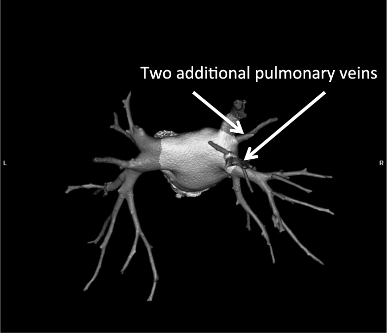
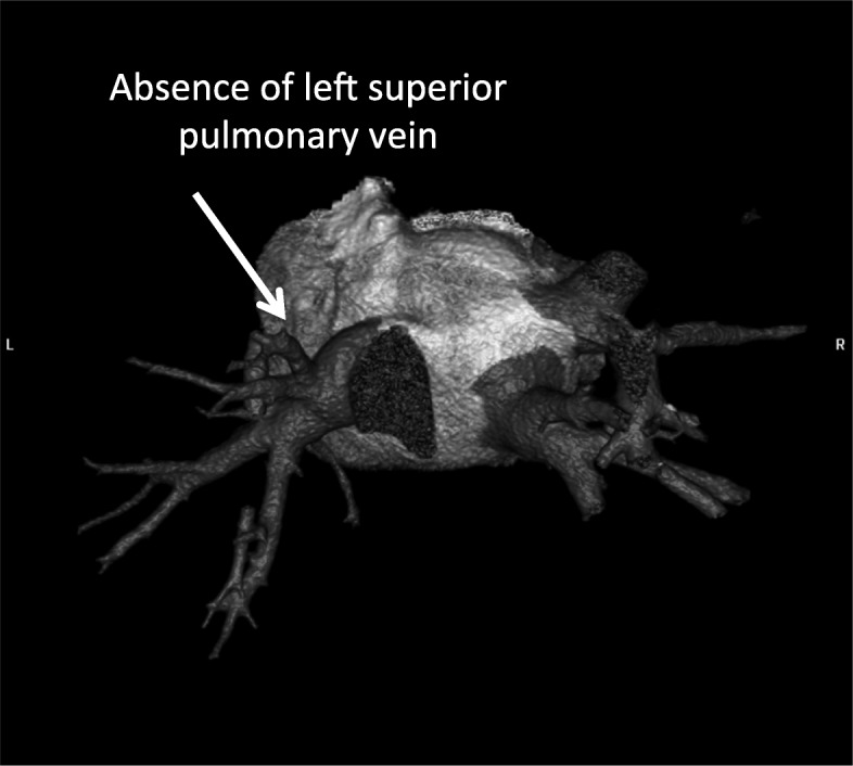
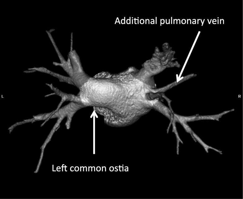
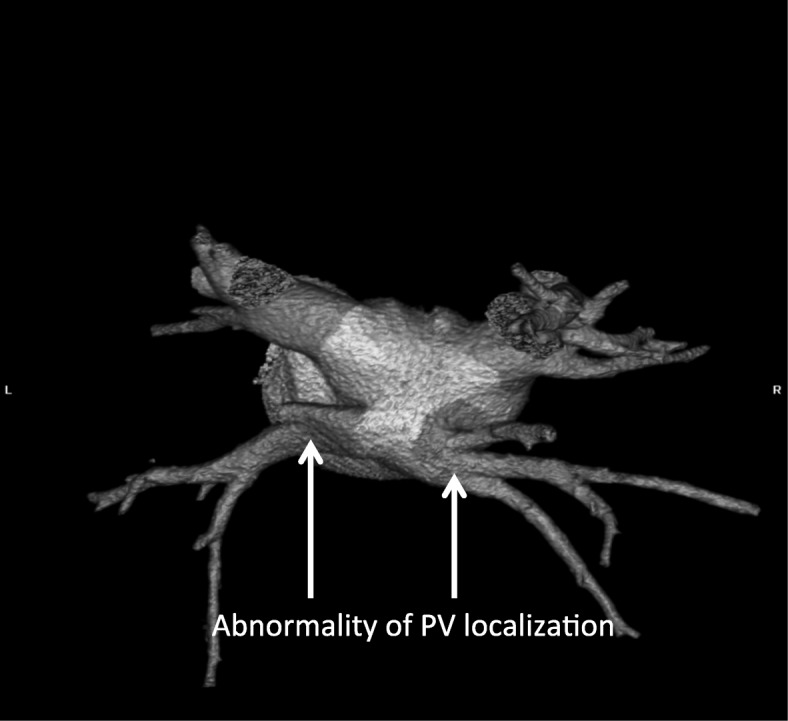
Results: Pulmonary veins anatomy variants were observed more frequently in the atrial fibrillation group - 83 pts. (37%) vs 6 pts. (15%) in CG; 9% (21 pts) left common ostia (CO), 2% (5 pts) right CO, 19% (42 pts) additional right PV (APV), (1.8%) 4 pts. APV left, 8% right early branching (EB) and 3.5% left EB. The LA diameter differed significantly in AF vs CG group (41.2 ± 6 mm vs 35 ± 4.2 mm, p < 0.0001) respectively.
Conclusions: The anomalies of pulmonary vein anatomy occurred more often in pts. with AF. They can be defined as an image biomarkers of atrial fibrillation. Right additional (middle) pulmonary vein was the most important anomaly detected in AF patients as well as enlargered diameters of the LA and PV ostia.
Keywords: Atrial fibrillation; CT angiography; Pulmonary vein isolation.
Conflict of interest statement
Ethics approval and consent to participate
The study was approved by the ethics committee (Medical University of Silesia ethics comittee) and conformed to the Declaration of Helsinki. An informed written consent was obtained from every patient enrolled in the study.
Consent for publication
n/a
Competing interests
The authors declare that they have no competing interests.
Publisher’s Note
Springer Nature remains neutral with regard to jurisdictional claims in published maps and institutional affiliations.
Figures
Similar articles
-
Comparison of pulmonary veins anatomy in patients with and without atrial fibrillation: analysis by multislice tomography.Int J Cardiol. 2011 Jan 21;146(2):181-5. doi: 10.1016/j.ijcard.2009.06.047. Epub 2009 Jul 25. Int J Cardiol. 2011. PMID: 19632731
-
Pulmonary vein anatomy and its variations in a Turkish atrial fibrillation cohort undergoing cryoballoon-based catheter ablation.Turk Kardiyol Dern Ars. 2017 Jan;45(1):42-48. doi: 10.5543/tkda.2016.12080. Turk Kardiyol Dern Ars. 2017. PMID: 28106019
-
Left atrial volume calculated by multi-detector computed tomography may predict successful pulmonary vein isolation in catheter ablation of atrial fibrillation.Europace. 2009 Oct;11(10):1289-94. doi: 10.1093/europace/eup198. Epub 2009 Jul 23. Europace. 2009. PMID: 19632980 Clinical Trial.
-
A meta-analysis of the relationship between anatomical variations of pulmonary veins and atrial fibrillation.Acta Cardiol. 2020 Feb;75(1):1-9. doi: 10.1080/00015385.2018.1544204. Epub 2019 Feb 8. Acta Cardiol. 2020. PMID: 30736723
-
Left atrial diverticula: Innocent bystanders or wolves in sheep's clothing?J Cardiovasc Electrophysiol. 2020 Sep;31(9):2484-2488. doi: 10.1111/jce.14581. Epub 2020 Jun 16. J Cardiovasc Electrophysiol. 2020. PMID: 32445428 Review.
Cited by
-
Silent cerebral ischemic lesions in ablation-naïve patients with non-valvular atrial fibrillation: Does the pulmonary vein anatomy matter?Cardiol J. 2025;32(1):35-42. doi: 10.5603/cj.99142. Epub 2024 Nov 26. Cardiol J. 2025. PMID: 39589069 Free PMC article.
-
Reduced Radiation Exposure Protocol during Computer Tomography of the Left Atrium Prior to Catheter Ablation in Patients with Atrial Fibrillation.Diagnostics (Basel). 2022 Mar 1;12(3):612. doi: 10.3390/diagnostics12030612. Diagnostics (Basel). 2022. PMID: 35328165 Free PMC article.
-
Morphological and morphometric study of pulmonary vein anatomy in relation to cardiac invasive and electrophysiological procedures.Anat Cell Biol. 2023 Dec 31;56(4):428-434. doi: 10.5115/acb.23.141. Epub 2023 Nov 10. Anat Cell Biol. 2023. PMID: 37946563 Free PMC article.
-
The Impact of Pulmonary Vein Anatomy on the Outcomes of Catheter Ablation for Atrial Fibrillation.Medicina (Kaunas). 2019 Nov 4;55(11):727. doi: 10.3390/medicina55110727. Medicina (Kaunas). 2019. PMID: 31690031 Free PMC article.
-
Linking statistical shape models and simulated function in the healthy adult human heart.PLoS Comput Biol. 2021 Apr 15;17(4):e1008851. doi: 10.1371/journal.pcbi.1008851. eCollection 2021 Apr. PLoS Comput Biol. 2021. PMID: 33857152 Free PMC article.
References
-
- Calkins H, Kuck KH, Cappato R et al. 2012 HRS/EHRA/EACS expert consensus statement on catheter and surgical ablation of atrial fibrillation: recommendations for patient selection, procedural techniques, patients management and follow-up, definitions, endpoints and research trial design: a report of the Heart Rhythm Society (HRS) task force on catheter and surgical ablation of atrial fibrillation. Heart Rhythm; 2012 9: 632–696. - PubMed
-
- Tekbas G, Ekici F, Tekbas E, et al. Evaluation of pulmonary vein variations in the middle pulmonary lobe with 64-slice multidetector computed tomography. Eur Rev Med Pharmacol Sci. 2011;15(12):1395–1400. - PubMed
MeSH terms
LinkOut - more resources
Full Text Sources
Other Literature Sources
Medical
