

Liprin-α1 modulates cancer cell signaling by transmembrane protein CD82 in adhesive membrane domains linked to cytoskeleton
- PMID: 30005669
- PMCID: PMC6045882
- DOI: 10.1186/s12964-018-0253-y
Liprin-α1 modulates cancer cell signaling by transmembrane protein CD82 in adhesive membrane domains linked to cytoskeleton
Abstract
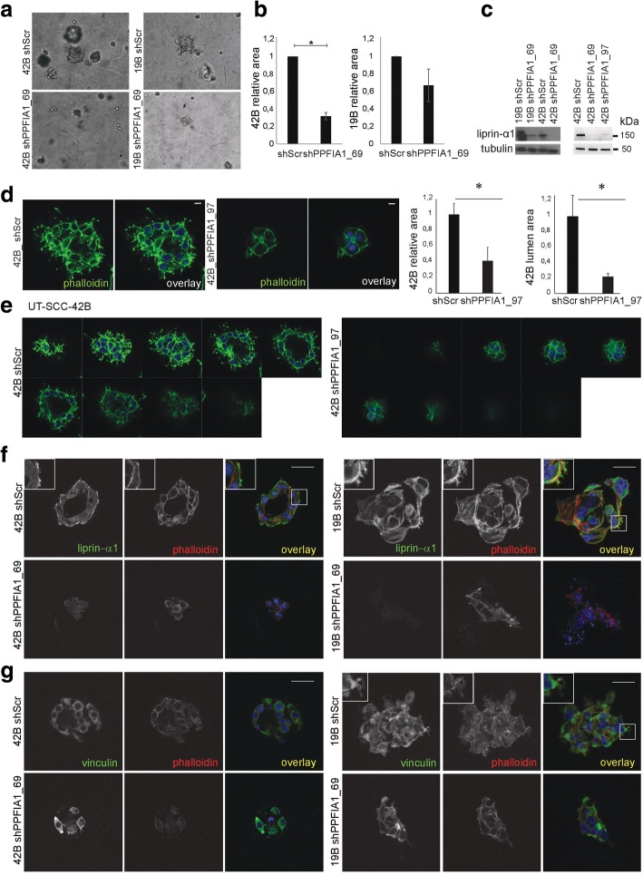
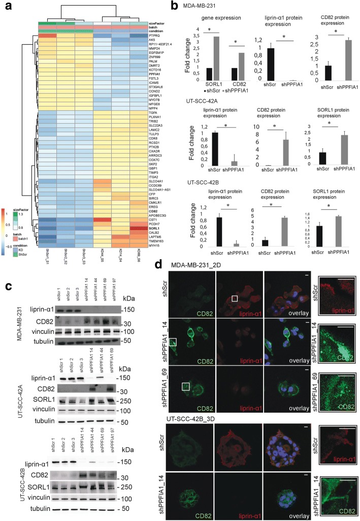
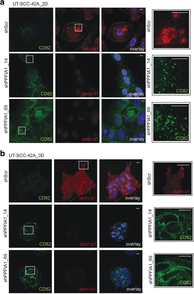
Background: PPFIA1 is located at the 11q13 region commonly amplified in cancer. The protein liprin-α1 encoded by PPF1A1 contributes to the adhesive and invasive structures of cytoskeletal elements and is located at the invadosomes in cancer cells. However, the precise mechanism of liprin-α1 function in cancer progression has remained elusive.
Methods: Invasion regulating activity of liprin-α1 was examined by analyzing the functions of squamous cell carcinoma of head and neck (HNSCC) cell lines in three-dimensional collagen I after RNAi mediated gene knockdown. Transcriptome profiling and Gene Set Enrichment Analysis from HNSCC and breast cancer cells were used to identify expression changes relevant to specific cellular localizations, biological processes and signaling pathways after PPFIA1 knockdown. The significance of the results was assessed by relevant statistical methods (Wald and Benjamini-Hochberg). Localization of proteins associated to liprin-α1 was studied by immunofluorescence in 2D and 3D conditions. The association of PPFIA1 amplification to HNSCC patient survival was explored using The Cancer Genome Atlas data.
Results: In this study, we show that liprin-α1 regulates biological processes related to membrane microdomains in breast carcinoma, as well as protein trafficking, cell-cell and cell-substrate contacts in HNSCC cell lines cultured in three-dimensional matrix. Importantly, we show that in all these cancer cells liprin-α1 knockdown leads to the upregulation of transmembrane protein CD82, which is a suppressor of metastasis in several solid tumors.
Conclusions: Our results provide novel information regarding the function of liprin-α1 in biological processes essential in cancer progression. The results reveal liprin-α1 as a novel regulator of CD82, linking liprin-α1 to the cancer cell invasion and metastasis pathways.
Keywords: Breast cancer; CD82; Head and neck cancer; Invasion; Liprin-α1; PPFIA1; RNA sequencing; Three-dimensional cell culture.
Conflict of interest statement
Ethics approval and consent to participate
Not applicable.
Consent for publication
Not applicable.
Competing interests
The authors declare that they have no competing interests.
Publisher’s Note
Springer Nature remains neutral with regard to jurisdictional claims in published maps and institutional affiliations.
Figures

Similar articles
-
Liprin-α1 contributes to oncogenic MAPK signaling by counteracting ERK activity.Mol Oncol. 2024 Mar;18(3):662-676. doi: 10.1002/1878-0261.13593. Epub 2024 Jan 24. Mol Oncol. 2024. PMID: 38264964 Free PMC article.
-
Liprin-α1 is a regulator of vimentin intermediate filament network in the cancer cell adhesion machinery.Sci Rep. 2016 Apr 14;6:24486. doi: 10.1038/srep24486. Sci Rep. 2016. PMID: 27075696 Free PMC article.
-
Amplification and overexpression of PPFIA1, a putative 11q13 invasion suppressor gene, in head and neck squamous cell carcinoma.Genes Chromosomes Cancer. 2008 Apr;47(4):353-62. doi: 10.1002/gcc.20539. Genes Chromosomes Cancer. 2008. PMID: 18196592
-
Role of Liprins in the Regulation of Tumor Cell Motility and Invasion.Curr Cancer Drug Targets. 2016;16(3):238-48. doi: 10.2174/156800961603160206124103. Curr Cancer Drug Targets. 2016. PMID: 26882029 Review.
-
Tetraspanin CD82: a suppressor of solid tumors and a modulator of membrane heterogeneity.Cancer Metastasis Rev. 2015 Dec;34(4):619-33. doi: 10.1007/s10555-015-9585-x. Cancer Metastasis Rev. 2015. PMID: 26335499 Review.
Cited by
-
Liprin-α1 contributes to oncogenic MAPK signaling by counteracting ERK activity.Mol Oncol. 2024 Mar;18(3):662-676. doi: 10.1002/1878-0261.13593. Epub 2024 Jan 24. Mol Oncol. 2024. PMID: 38264964 Free PMC article.
-
Two circPPFIA1s negatively regulate liver metastasis of colon cancer via miR-155-5p/CDX1 and HuR/RAB36.Mol Cancer. 2022 Oct 12;21(1):197. doi: 10.1186/s12943-022-01667-w. Mol Cancer. 2022. PMID: 36224588 Free PMC article.
-
PPFIA1 expression associates with poor response to endocrine treatment in luminal breast cancer.BMC Cancer. 2020 May 14;20(1):425. doi: 10.1186/s12885-020-06939-6. BMC Cancer. 2020. PMID: 32410585 Free PMC article.
-
The Structural Dynamics, Complexity of Interactions, and Functions in Cancer of Multi-SAM Containing Proteins.Cancers (Basel). 2023 Jun 1;15(11):3019. doi: 10.3390/cancers15113019. Cancers (Basel). 2023. PMID: 37296980 Free PMC article. Review.
-
Copy number alterations identify a smoking-associated expression signature predictive of poor outcome in head and neck squamous cell carcinoma.Cancer Genet. 2021 Aug;256-257:136-148. doi: 10.1016/j.cancergen.2021.05.011. Epub 2021 May 28. Cancer Genet. 2021. PMID: 34130230 Free PMC article.
References
-
- Jarvinen AK, Autio R, Haapa-Paananen S, Wolf M, Saarela M, Grenman R, Leivo I, Kallioniemi O, Makitie AA, Monni O. Identification of target genes in laryngeal squamous cell carcinoma by high-resolution copy number and gene expression microarray analyses. Oncogene. 2006;25(52):6997–7008. doi: 10.1038/sj.onc.1209690. - DOI - PubMed
-
- Meredith SD, Levine PA, Burns JA, Gaffey MJ, Boyd JC, Weiss LM, Erickson NL, Williams ME. Chromosome 11q13 amplification in head and neck squamous cell carcinoma. Association with poor prognosis. Arch Otolaryngol Head Neck Surg. 1995;121(7):790–794. doi: 10.1001/archotol.1995.01890070076016. - DOI - PubMed
-
- Akervall JA, Jin Y, Wennerberg JP, Zatterstrom UK, Kjellen E, Mertens F, Willen R, Mandahl N, Heim S, Mitelman F. Chromosomal abnormalities involving 11q13 are associated with poor prognosis in patients with squamous cell carcinoma of the head and neck. Cancer. 1995;76(5):853–859. doi: 10.1002/1097-0142(19950901)76:5<853::AID-CNCR2820760520>3.0.CO;2-6. - DOI - PubMed
-
- Schuuring E, Verhoeven E, Vantinteren H, Peterse JL, Nunnink B, Thunnissen FBJM, Devilee P, Cornelisse CJ, Vandevijver MJ, Mooi WJ, et al. Amplification of genes within the chromosome-11q13 region is indicative of poor prognosis in patients with operable breast-Cancer. Cancer Res. 1992;52(19):5229–5234. - PubMed
Publication types
MeSH terms
Substances
LinkOut - more resources
Full Text Sources
Other Literature Sources
Molecular Biology Databases
Miscellaneous
