Pyeloduodenal Fistula in Xanthogranulomatous Pyelonephritis: A Series of Two Cases
- PMID: 30005725
- PMCID: PMC6045498
- DOI: 10.7812/TPP/17-150
Pyeloduodenal Fistula in Xanthogranulomatous Pyelonephritis: A Series of Two Cases
Abstract
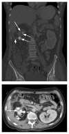
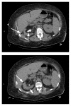
Xanthogranulomatous inflammation, characterized by destruction and replacement of tissues with chronic inflammatory cells, including foamy histiocytes and hemosiderin-laden macrophages, is uncommon. In patients with xanthogranulomatous pyelonephritis, inflammation may extend from the kidney to the overlying duodenum, creating a pyeloduodenal fistula that further complicates medical and surgical management. We present two cases with recurrent kidney infections who each ultimately received a nephrectomy and repair of their duodenal fistula.
Conflict of interest statement
The author(s) have no conflicts of interest to disclose.
Figures


References
-
- Hode E, Josse C, Mechaouri M, Garnier L, Verhaeghe P. [Pyeloduodenal fistula. Apropos of a new case]. [Article in French] J Chir (Paris) 1990 May;127(5):281–5. - PubMed
-
- Dahami Z, Dakir M, Aboutaieb R, et al. [Diffuse xanthogranulomatous pyelonephritis: Clinical, anatomopathologic, and therapeutic features. Report ot 9 cases and review of the literature]. [Article in French] Ann Urol (Paris) 2001 Nov;35(6):309–14. - PubMed
-
- Parsons MA, Harris SC, Longstaff AJ, Grainger RG. Xanthogranulomatous pyelonephritis: A pathological, clinical and aetiological analysis of 87 cases. Diagn Histopathol. 1983 Jul-Dec;6(3–4):203–19. - PubMed
-
- Sallami S, Rhouma SB, Rebai S, et al. Spontaneous pyeloduodenal fistula complicating a xanthogranulomatous pyelonephritis. Ibnosina Journal of Medicine and Biomedical Sciences. 2010;2(6):283–7. doi: 10.4103/1947-489X.211009. - DOI
Publication types
MeSH terms
LinkOut - more resources
Full Text Sources
Other Literature Sources
Medical

