. 2018 Dec;24(12):2397-2408.
doi: 10.1016/j.bbmt.2018.07.003. Epub 2018 Jul 10.

Host-Derived Serine Protease Inhibitor 6 Provides Granzyme B-Independent Protection of Intestinal Epithelial Cells in Murine Graft-versus-Host Disease

Affiliations

Host-Derived Serine Protease Inhibitor 6 Provides Granzyme B-Independent Protection of Intestinal Epithelial Cells in Murine Graft-versus-Host Disease

Hemn Mohammadpour et al. Biol Blood Marrow Transplant. 2018 Dec.

Abstract

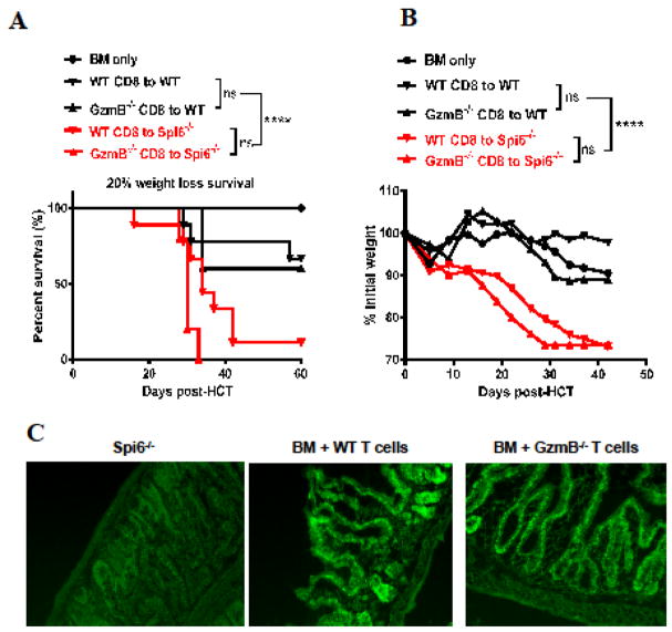
Graft-versus-host disease (GVHD) is a serious complication after allogeneic hematopoietic cell transplantation (allo-HCT) that limits the therapeutic potential of this treatment. Host antigen-presenting cells (APCs) play a vital role in activating donor T cells that subsequently use granzyme B (GzmB) and other cytotoxic molecules to damage host normal tissues. Serine protease inhibitor 6 (Spi6), known as the sole endogenous inhibitor of GzmB, has been implicated in protecting T cells and APCs against GzmB-inflicted damage. In this study we used murine models to examine the previously unknown role of host-derived Spi6 in GVHD pathogenesis. Our results indicated that host Spi6 deficiency exacerbated GVHD as evidenced by significantly increased lethality and clinical and histopathologic scores. Using bone marrow chimera system, we found that Spi6 in nonhematopoietic tissue played a dominant role in protecting against GVHD and was significantly upregulated in intestinal epithelial cells after allo-HCT, whereas Spi6 in hematopoietic APCs surprisingly suppressed alloreactive T cell response. Interestingly, the protective effect of Spi6 and its expression in intestinal epithelial cells appeared to be independent of donor-derived GzmB. We used in silico modeling to explore potential targets of Spi6. Interaction tested in silico demonstrated that Spi6 could inhibit caspase-3 and caspase-8 with the same functional loop that inhibits GzmB but was not capable of forming stable interaction with caspase-1 or granzyme A. Using an in vitro co-culture system, we further identified that donor T cell-derived IFN-γ was important for inducing Spi6 expression in an intestinal epithelial cell line. Altogether, our data indicate that host Spi6 plays a novel, GzmB-independent role in regulating alloreactive T cell response and protecting intestinal epithelial cells. Therefore, enhancing host-derived Spi6 function has the potential to reduce GVHD.

Keywords: Allogenic transplantation; GVHD; Granzyme B; Spi6.


Conflict of interest statement

Disclosure of Conflicts of Interest

The authors have no special/competing interests to disclose.

Figures

Figure 1
Figure 1. Spi6−/− hosts exhibit increased lethal and clinical GVHD
(A, B and C) WT or Spi6−/− host (H-2b) were lethally irradiated with 1100 cGy at day -1. At day 0, mice were injected with 6x10⁶ BM cells alone or combined with 5x10⁶ Pan T cells isolated from BALB/cJ (H-2d) donor mice. Host survival (A), body weight (B), and clinical score (C) were monitored after allo-HCT. Combined data from two independent experiments are presented (n=5 for control and n=15 for experimental groups). (D, E and F) WT or Spi6−/− host (H-2b) were lethally irradiated with 1100 rad at day -1. At day 0, mice were injected with 6x10⁶ BM cells alone or combined with 3x10⁶ CD8+ T cells isolated from BALB/cJ (H-2d) donor mice. Host survival (D), body weight (E), and clinical score (F) were monitored after allo-HCT. Results are presented from one individual experiment (n=2 for control and n=6 for experimental groups) (G, H and I) WT or Spi6−/− host (H-2b) were lethally irradiated with 1100rad at day -1. At day 0, mice were injected with 6x10⁶ BM cells alone or combined with 2.2x10⁶ CD8+ T cells isolated from 129/SvJ (H-2b) donor mice. Host survival (G), body weight (H), and clinical score (I) were monitored after allo-HCT. Combined data from two independent experiments are presented (n=6 for control and n=15 for experimental groups). Lethality condition was determined when either actual death occurred (A–C) or when hosts lost >20% body weight (D–I). Of note, in the 129/SvJ to B6 model (D–I) in which 2.2 × 10⁶ CD8+ T cells did not cause actual lethality to recipient mice, survival curves were determined by 20% weight loss combined with other criteria such as activity, severity of diarrhea and posture changes after consulting with veterinarian from animal facility. Statistical significances for Kaplan-Meier survival curves are evaluated by Log-rank (Mantel-Cox) tests and body weight loss, and clinical GVHD scores are evaluated by two-way ANOVA. P values are presented to compare WT versus Spi6−/− host after BM + T cells allo-HCT for all graphs.
Figure 2
Figure 2. Spi6−/− hosts exhibit increased histopathological GVHD and GI tract permeability
(A) WT or Spi6−/− host (H-2b) were lethally irradiated with 1100 cGy at day -1. At day 0, mice were injected with 6x10⁶ BM cells alone or combined with 2.2x10⁶ CD8+ T cells isolated from 129/SvJ (H-2b) donor mice. At day 60, all of the host mice were sacrificed for histopathological analysis (n=5; magnification is 100x). GVHD scores of large intestine, small intestine, and liver are shown in the summary graphs. (B and C) WT or Spi6−/− host (H-2b) were lethally irradiated with 1100 rad at day -1. At day 0, mice were injected with 6x10⁶ BM cells alone or combined with 3x10⁶ from either Pan T cells (B) or CD8+ T cells (C) isolated from BALB/cJ (H-2d) donor mice. At day 6 (B) or day 22 (C), FITC-dextran GI tract permeability tests were performed as described in materials and methods (N=5). One-way ANOVA test was performed for statistical significance. ** P<0.01; * P<0.05.
Figure 3
Figure 3. Spi6 in non-hematopoietic tissue plays a dominant role in protecting against GVHD and is upregulated in intestinal epithelial cells after allo-HCT
(A) Chimeras were made between C57BL/6 WT and Spi6−/− hosts with syngeneic transplantation of 5x10⁶ BM plus 5x10⁶ splenocytes. After 3 months, the chimeric hosts were lethally irradiated with 800 cGy and injected with 6x10⁶ BM alone or combined with 2.5x10⁶ CD8+ T cells harvested from 129/SvJ donors. Statistical significances for Kaplan-Meier survival curves were evaluated by Log-rank (Mantel-Cox) tests (combined from two experiments and 5–8 mice per experiments). *** P<0.001; ** P<0.01. (B–D) Lethally irradiated WT and Spi6−/− C57BL/B6 (H-2b) mice were transplanted with 6x10⁶ BM cells alone or combined with 3x10⁶ CD8+ T cells from MHC-mismatched BALB/cJ on day 0. At day 14, small intestines were isolated. Immunofluorescence staining was performed on (B) naïve small intestine, (C) BM only and (D) BM plus T cells. Magnification is 100x. Spi6 expression on green (Rabbit anti-mouse Spi6 as primary antibody (Hycultbiotech) and Alexa Flour 488 donkey to rabbit IgG (Abcam) as secondary antibody) and LGR5 (intestinal stem cells marker; goat polyclonal IgG LGR-5 (C-16) as primary antibody and Alexa Flour 568 donkey to goat IgG (Abcam) as secondary antibody) expression on red color are presented. (F) Spi6 expression were quantified by green color mean fluorescence intensity (MFI) using ImageJ software.
Figure 4
Figure 4. Spi6 in APCs suppresses T cell proliferation in vitro and Spi6−/− hosts exhibit increased T cell proliferation and activation in vivo
(A) MLR was performed using C57BL/6 WT or Spi6−/− splenocytes (3000 cGy irradiation) as stimulators and BALB/c Pant T cells as responders (CFSE labeled) with a ratio of 3:1. After co-culture for 4 days, cell proliferation analyzed by flow cytometry for CFSE dilution. Histogram plots are gated on Live/Dead–H2d+ TCRβ+ CD4+ or CD8+ T cells. (B) C57BL/6 WT or Spi6−/− mice received 1100 cGy irradiation on day -1 and were subsequently transplanted on day 0 with 6x10⁶ BM + 5x10⁶ PanT cells. At day 6, absolute number were calculated based on Live TCRβ+ H-2d+ CD4+ or CD8+ and total cell numbers. (C) C57BL/6 WT or Spi6−/− mice received 1100 cGy irradiation on day -1 and were subsequently transplanted on day 0 with 6x10⁶ BM + 5x10⁶ Ef670 labeled PanT from BALB/cJ donors. Ef670 dilution was assessed in live TCRβ+ H-2d+CD4+ or TCRβ+H-2d+CD8+ T cells. Summary data presented from 10 mice of each group at day 6 after allo-HCT. (D, E and F) C57BL/6 WT or Spi6−/− hosts were irradiated with 1100 cGy at day -1. At day 0, mice were injected with 6x10⁶ BM cells alone or combined with (D) 3x10⁶ Pan T or (E) 3x10⁶ CD8+ T cells isolated from WT BALB/cJ (H-2d) donor mice. (D) Effector cells gated on live H-2d+CD4+ cells. (E) Activated CD8+CD122+cells gated from live H-2d+ cells. (F) Summary data presented from 10 mice of each group at day 6 after allo-HCT for both D and E respectively. Student’s t test was performed for statistical significance. *** P<0.001; ** P<0.01; * P<0.05.
Figure 5
Figure 5. Host Spi6-mediated protection is independent of donor T cell-derived GzmB
(A and B) WT or Spi6−/− host (H-2b) were lethally irradiated with 1100 cGy at day -1. At day 0, mice were injected with 6x10⁶ BM cells alone or combined with 2.2x10⁶ CD8+ T cells isolated from WT or Gzm−/− 129/SvJ (H-2b) donor mice. Host survival (A) and body weight (B) were monitored after allo-HCT (BM only (n=5), WT CD8 to WT and GzmB−/− CD8 to WT (n=9), WT CD8 to Spi6−/− and GzmB−/− CD8 to Spi6−/− (n=5)). Statistical significances for Kaplan-Meier survival curves are evaluated by Log-rank (Mantel-Cox) tests and body weight loss was evaluated by two-way ANOVA. **** P < 0.0001 (C) Lethally irradiated WT and Spi6−/− C57BL/B6 (H-2b) mice were transplanted with 6x10⁶ BM cells alone or combined with 2.2x10⁶ CD8+ T cells from WT and GzmB−/− 129/SvJ on day 0. At day 14, small intestines were isolated. Immunofluorescence staining of Spi6 expression was performed on naïve small intestine, BM only, and BM plus T cells. Magnification is 100x.
Figure 6
Figure 6. Spi6 interacts with Granzyme B, caspase-3 and caspase-8 with common active loop
The 3D structure refined model of the Spi6 (blue) and its interaction with (A) GzmB, (B) caspase-3 and (C) caspase-8, all in green color. Potential interacted amino acid in Spi6, GzmB, caspase-3 and caspase-8 after interaction is colored in red. (D) The results of docking analyses along with the binding energies.
Figure 7
Figure 7. T cell-derived IFN-γ induces Spi6 expression on intestinal epithelial cells
MODE-K cell line was incubated with either CD8+ T cells or CD4+ T cells with or without CD28 for 48 hours. Expressions of Spi6, GzmB, IFN-γ and TNF-α were examined with flow cytometry. (A) Representative flow cytometry plot (B) and statistical analysis of GzmB, IFN-γ and TNF-α expression by either CD8+ T cells or CD4+ T cells after co-culture with MODE-K cell line. (C) Spi6 expression in MODE-K cells after co-culture with either CD8+ T cells or CD4+ T cells with or without CD28 for 48 hours. (D- E) MODE-K cells co-cultured with recombinant mouse IFN-γ (20ng/ml) or TNF-α (20ng/ml) for 48 hours and Spi6 expression checked with flow cytometry. (F-G) MODE-K cell line were incubated with CD4+ T cells with CD28 in the presence of either anti-IFN-γ antibody (53μ/ml) or isotype control antibody (53μ/ml) for 48 hours and Spi6 expression checked with flow cytometry. One-way ANOVA test was performed for statistical significance ***P<0.001; **P<0.01; *P<0.05. Data are presented from three independent experiments.

References

    1. Ramadan A, Paczesny S. Various forms of tissue damage and danger signals following hematopoietic stem-cell transplantation. Frontiers in immunology. 2015:6. - PMC - PubMed
    1. Shlomchik WD, Couzens MS, Tang CB, et al. Prevention of graft versus host disease by inactivation of host antigen-presenting cells. Science. 1999;285:412–415. - PubMed
    1. Matte CC, Liu J, Cormier J, et al. Donor APCs are required for maximal GVHD but not for GVL. Nature medicine. 2004;10:987. - PubMed
    1. Highfill SL, Rodriguez PC, Zhou Q, et al. Bone marrow myeloid-derived suppressor cells (MDSCs) inhibit graft-versus-host disease (GVHD) via an arginase-1–dependent mechanism that is up-regulated by interleukin-13. Blood. 2010;116:5738–5747. - PMC - PubMed
    1. Ghosh A, Smith M, James SE, et al. Donor CD19-CAR T Cells Exert Potent Graft-versus-Lymphoma Activity With Diminished Graft-versus-Host Activity. Nature medicine. 2017;23:242. - PMC - PubMed

Publication types