. 2018 Aug 30;8(9):2979-2990.
doi: 10.1534/g3.118.200572.

CORL Expression and Function in Insulin Producing Neurons Reversibly Influences Adult Longevity in Drosophila

Affiliations

CORL Expression and Function in Insulin Producing Neurons Reversibly Influences Adult Longevity in Drosophila

Nancy L Tran et al. G3 (Bethesda). .

Abstract

CORL proteins (known as SKOR in mice, Fussel in humans and fussel in Flybase) are a family of CNS specific proteins related to Sno/Ski oncogenes. Their developmental and adult roles are largely unknown. A Drosophila CORL (dCORL) reporter gene is expressed in all Drosophila insulin-like peptide 2 (dILP2) neurons of the pars intercerebralis (PI) of the larval and adult brain. The transcription factor Drifter is also expressed in the PI in a subset of dCORL and dILP2 expressing neurons and in several non-dILP2 neurons. dCORL mutant virgin adult brains are missing all dILP2 neurons that do not also express Drifter. This phenotype is also seen when expressing dCORL-RNAi in neurosecretory cells of the PI. dCORL mutant virgin adults of both sexes have a significantly shorter lifespan than their parental strain. This longevity defect is completely reversed by mating (lifespan increases over 50% for males and females). Analyses of dCORL mutant mated adult brains revealed a complete rescue of dILP2 neurons without Drifter. Taken together, the data suggest that dCORL participates in a neural network connecting the insulin signaling pathway, longevity and mating. The conserved sequence and CNS specificity of all CORL proteins imply that this network may be operating in mammals.

Keywords: Drifter; Fussel/SKOR; dILP2; lifespan extension; pars intercerebralis.


Figures

Figure 1
Figure 1
dCORL is expressed in all dILP2 neurons of the larval brain. Dorsal view of AH.lacZ transgenic larval female brains with anterior up. A) AH.lacZ (green-nuclear), Fas2 (blue–marks a distinct subset of neurons including the optic lobe) and Drifter (red-nuclear) at 20X. AH.lacZ is a nuclear-lacZ reporter containing genomic DNA upstream of dCORL. Expression of both lacZ and Drifter in the PI is seen. B) 40X stack and single slice of the PI in the left hemisphere from the same brain. Co-expression of AH.lacZ and Drifter in a subset of neurons is visible. C) AH.lacZ (green), Fas2 (blue) and dILP2 (red-cytoplasmic) at 20X. Coexpression of AH.lacZ and dILP2 in the PI is evident (neurons with red cytoplasm have green nuclei). Axons from the coexpressing neurons in both hemispheres extend medially. D) 40X stack and single slice of the PI in the left hemisphere from the same brain. Coexpression of AH.lacZ and dILP2 in all dILP2 neurons of the PI is confirmed (all neurons with red cytoplasm have green nuclei). An additional AH.lacZ neuron sits just medial to the co-expressing neurons. Note that the bottom four panels in Fig. 5 of Tran et al. (2018) are intentionally similar to the bottom four panels here. The point of the similarity is to document the logic connecting these papers.
Figure 2
Figure 2
dCORL is expressed in all dILP2 neurons of the adult brain. One day old AH.lacZ transgenic virgin adult female brains in dorsal view with anterior up. A) AH.lacZ (green), Fas2 (blue marks mushroom body lobes employed to determine orientation) and Toy (red-nuclear) at 20X. AH.lacZ expression is clearly visible in the PI. A’) 40X views in 3-color and as single channels. An arbitrary line in the 3-color view surrounding the PI serves as a landmark in the individual channels. Strong AH.lacZ expression in neurons of the PI overflows the nucleus into the descending axon bundle. A few scattered neurons in the posterior display AH.lacZ expression, as do 2-3 neurons at the border of the optic lobe. No coexpression between AH.lacZ and Toy in the PI is noted. Toy expressing neurons are located ventral to AH.lacZ neurons and their nuclei are a different size. B) AH.lacZ (green), Fas2 (blue) and Elav (red-nuclear) show coexpression (yellow) in neurons of the PI. C) AH.lacZ (green), Toy (blue) and Repo (red-nuclear) shows no coexpression in glia of the PI (red and green neurons are adjacent). D) AH.lacZ (green), Fas2 (blue) and Drifter (red) shows coexpression of AH.lacZ and Drifter (yellow) in a subset of neurons within the PI. E) AH.lacZ (green), Fas2 (blue) and dILP2 (red) shows coexpression in all dILP2 neurons of the PI (all neurons with red cytoplasm have green nuclei). An additional AH.lacZ neuron without dILP2 is also visible in the PI of each hemisphere, sitting medially.
Figure 3
Figure 3
dCORL mutant larval brains are missing all dILP2 neurons lacking Drifter. Dorsal view of larval female brains with anterior up stained with Drifter (green - nuclear), Fas2 (blue) and dILP2 (red - cytoplasmic). A) Wild type (y1w67c23) at 20X shows the presence of dILP2 and Drifter in the PI. B) 40X stack and single slice of the PI of the left hemisphere from the same brain. The single slice view shows that the dILP2 neurons are essentially in a monolayer (all 8 neurons are visible in a single slice). There are neurons that express Drifter alone, three neurons expressing dILP2 alone (white arrowheads), and five neurons that express both. C) Df(4)dCORL brain (larvae was aged to the same developmental point as wild type before dissection) at 20X shown to scale. Notwithstanding the complete disarray of Fas2 and Drifter staining as well as the statistically significant reduction in Df(4)dCORL brain size, both dILP2 and Drifter are present in the PI. D) 40X stack and single slice of the PI of the left hemisphere from the same brain. The single slice view shows that there are just 5 dILP2 neurons (vs. 8 in wild type) and that their topology is altered. These are no longer in a monolayer (only 3 neurons are visible in a single slice). There are no neurons expressing dILP2 alone as all 5 dILP2 neurons express Drifter.
Figure 4
Figure 4
dCORL mutant virgin adult brains are missing all dILP2 neurons lacking Drifter. One day old virgin adult female brain in dorsal view with anterior up displaying Drifter (green), Fas2 (blue) and dILP2 (red). A) Wild type (y1w67c23) at 20X shows dILP2 and Drifter in the PI. B) 40X stack of the PI from the same brain: (top) 2-color, (middle and bottom) single channel of dILP2 (red) and Drifter (green). The 2-color view shows that dILP2-Drifter coexpressing neurons form a single straight row along the apical surface of the PI with additional dILP2 neurons forming an inverted triangle. There are neurons that express Drifter alone in the apical row. C) Three slices show the pyramidal structure of the PI (not all dILP2 neurons are visible in the same slice). Four dILP2 neurons that do not express Drifter are indicated (white arrowheads). D) Df(4)dCORL brain at 20X shown to scale with dILP2 and Drifter in the PI. E) 40X stack of the PI from the same brain: (top) 2-color, (middle and bottom) single channel of dILP2 (red) and Drifter (green). The 2-color view shows there has been a decrease in the number of dILP2 expressing neurons. The dILP2-Drifter coexpressing neurons still form a single row but the absence of additional dILP2 neurons medially caused the apical row to collapse into a V-shape. There are neurons that express Drifter alone in the dILP2-Drifter V-shape and their number has not changed. F) Three slices show that the V-shaped row of dILP2-Drifter neurons has a non-wild type structure. No dILP2 neurons that do not express Drifter are present.
Figure 5
Figure 5
dCORL mutant virgin adult brains are missing Dac neurons in the PI. One day old virgin adult female brains in dorsal view with anterior up stained with Drifter (green), Dac (blue - nuclear) and dILP2 (red). A) Wild type (y1w67c23) at 20X shows the presence of all proteins in the PI. B) 40X stack of the PI from the same brain: (top) 2-color view, (middle and bottom) single channel views of dILP2 (red) and Dac (blue). The 2-color view shows dILP2-Dac coexpressing neurons are surrounded by many Dac neurons that form an inverted triangle. C) Three slices covering 8 microns show that all dILP2 neurons express Dac. D) Df(4)dCORL brain at 20X shown to scale. All proteins are present in the PI. E) 40X stack of the PI from the same brain: (top) 2-color view, (middle and bottom) single channel views of dILP2 (red) and Dac (blue). The 2-color view shows that there has been a modest decrease in the number of dILP2-Dac coexpressing neurons and a substantial decrease in Dac neurons. The dILP2-Dac coexpressing neurons and the majority of remaining Dac neurons form a single row along the apical surface of the PI (with the exception of a single dILP2 cell). The absence of additional dILP2-Dac and Dac only neurons medially has caused the apical row to collapse into a V-shape. F) Three slices covering a span of 8 microns shows that all dILP2 neurons express Dac.
Figure 6
Figure 6
dCORL-RNAi in the PI phenocopies the Df(4)dCORL dILP2 phenotype. One day old virgin adult female brains in dorsal view with anterior up. A) Wild type (y1w67c23) with OK107.Gal4 driving UAS.lacZ at 20X displaying lacZ (green), Fas2 (blue) and dILP2 (red) shows the presence of lacZ with Fas2 in the mushroom body lobes and lacZ with dILP2 in the PI. B) 40X stack of the PI from the same brain: (top) 2-color view, (middle) single channel view of dILP2 (red). Both views show the wild type pyramidal structure of lacZ and dILP2 expressing neurons in the PI. C) One slice showing the same topology. D) Wild type (y1w67c23) with OK107.Gal4 driving UAS.dCORL-RNAi displaying Drifter (green), Fas2 (blue) and dILP2 (red). Mushroom body defects due to loss of dCORL in Df(4)dCORL adult brains are visible that verify dCORL-RNAi expression. E) 40X stack of the PI from the same brain: (top) 2-color view, (middle and bottom) single channel views of dILP2 (red) and Drifter (green). These views show that there has been a modest decrease in the number of dILP2 expressing neurons and that their topology has flattened out though not to the extent seen in Df(4)dCORL. F) One slice shows that all dILP2 neurons express Drifter, a phenocopy of the Df(4)dCORL dILP2 phenotype.
Figure 7
Figure 7
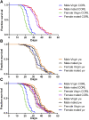
dCORL mutant virgin adult longevity defects are fully rescued by mating in both sexes. Cohorts of 100 flies of each genotype and mating status were analyzed in parallel under identical conditions. Mortality assessments were made daily. Numerical data from this experiment is in Tables 1 and 2 with a much larger set of genotypic controls shown in Table S1. Note that Df(4)dCORL has a yw background. A) Df(4)dCORL virgins display a relatively short lifespan but upon mating there is a highly significant extension of lifespan for both sexes. B) yw virgins and mated flies of both sexes show no lifespan differences. C) Df(4)dCORL virgins of both sexes show a highly significant lifespan reduction vs. yw. Upon mating, the lifespan reduction of Df(4)dCORL virgins is completely rescued as mated Df(4)dCORL adults of both sexes are not different from yw.
Figure 8
Figure 8
dILP2 neurons lacking Drifter are fully rescued by mating in dCORL mutant adult brains. Df(4)dCORL mated adult female brains shown as in Fig. 4. A) Three day old at 20X shows rescue of PI topology compared to dCORL mutant virgin females. B) 40X stack of the PI from the same brain: (top) 2-color view, (middle and bottom) single channel views of dILP2 (red) and Drifter (green). Three day old mated females display an increase in dILP2 expressing neurons vs. virgin females due to the presence of rescued neurons expressing dILP2 only. C) Three slices at 40X covering 10 microns show the pyramidal structure of the PI is similar to wild type though more compact. Multiple dILP2 neurons lacking Drifter are present (white arrowheads). D) Fifteen day old at 20X shows that the rescue of PI topology persists. E) 40X stack of the PI from the same brain: (top) 2-color view, (middle and bottom) single channel views of dILP2 (red) and Drifter (green). Fifteen day old mated female also displays an increase in dILP2 expressing neurons vs. virgins indicating that rescued neurons expressing dILP2 only persist. F) Three slices covering a span of 24 microns show the pyramidal structure of the PI is similar to wild type though more expansive. Multiple rescued dILP2 neurons lacking Drifter (white arrowheads) are present after two weeks.

Similar articles

Cited by

References

    1. Bai H., Kang P., Hernandez A. M., Tatar M., 2013. Activin signaling targeted by insulin/dFOXO regulates aging and muscle proteostasis in Drosophila. PLoS Genet. 9: e1003941 10.1371/journal.pgen.1003941 - DOI - PMC - PubMed
    1. Bogdanik L., Mohrmann R., Ramaekers A., Bockaert J., Grau Y., et al. , 2004. The Drosophila metabotropic glutamate receptor DmGluRA regulates activity-dependent synaptic facilitation and fine synaptic morphology. J. Neurosci. 24: 9105–9116. 10.1523/JNEUROSCI.2724-04.2004 - DOI - PMC - PubMed
    1. Bowman E., Tatar M., 2016. Reproduction regulates Drosophila nutrient intake through independent effects of egg production and sex peptide: Implications for aging. Nutr. Healthy Aging 4: 55–61. 10.3233/NHA-1613 - DOI - PMC - PubMed
    1. Broughton S., Piper M., Ikeya T., Bass T., Jacobson J., et al. , 2005. Longer lifespan, altered metabolism, and stress resistance in Drosophila from ablation of cells making insulin-like ligands. Proc. Natl. Acad. Sci. USA 102: 3105–3110. 10.1073/pnas.0405775102 - DOI - PMC - PubMed
    1. Chapman T., Liddle L., Kalb J., Wolfner M., Partridge L., 1995. Cost of mating in Drosophila melanogaster females is mediated by male accessory gland products. Nature 373: 241–244. - PubMed

Publication types

LinkOut - more resources