

Comment
doi: 10.1083/jcb.201807036.
Epub 2018 Jul 13.
A new look for the growing microtubule end?
Affiliations
- PMID: 30006463
- PMCID: PMC6080924
- DOI: 10.1083/jcb.201807036
Item in Clipboard
Comment
A new look for the growing microtubule end?
J Cell Biol.
.
Abstract
What does the end of a growing microtubule look like? In this issue, McIntosh et al. (2018. J. Cell Biol. https://doi.org/10.1083/jcb.201802138) use electron tomography to provide state-of-the-art three-dimensional images of microtubule ends in cells and in vitro, yielding an unexpected answer to this fundamental question.
© 2018 Rice.
Figures
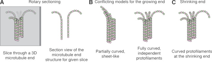
Illustration of rotary sectioning and the different views of microtubule end structure. (A) Rotary sectioning. Left: Slices through 3D tomographs of microtubule ends are taken at various angles. The αβ-tubulin subunits of the microtubule are represented as pink and green circles, and the gray plane represents one such slice through the volume. Bright subunits are in front of the plane, faint subunits are behind it, and intermediate shaded subunits are in the slice. Right: View of the resulting slice, showing the end structure. The vertical head-to-tail assemblies of αβ-tubulin are called protofilaments. (B) Conflicting models for how microtubules grow. Left: Sheet-like, partially curved extensions on a subset of protofilaments. Right: All protofilaments elongate independently and are fully curved. (C) Cartoon of a shrinking microtubule end, with the ends of protofilaments fully curved.
Comment on
-
Microtubules grow by the addition of bent guanosine triphosphate tubulin to the tips of curved protofilaments.J Cell Biol. 2018 Aug 6;217(8):2691-2708. doi: 10.1083/jcb.201802138. Epub 2018 May 23. J Cell Biol. 2018. PMID: 29794031 Free PMC article.
References
-
- Atherton J., Jiang K., Stangier M.M., Luo Y., Hua S., Houben K., van Hooff J.J.E., Joseph A.-P., Scarabelli G., Grant B.J., et al. 2017. A structural model for microtubule minus-end recognition and protection by CAMSAP proteins. Nat. Struct. Mol. Biol. 24:931–943. 10.1038/nsmb.3483 - DOI - PMC - PubMed
Publication types
MeSH terms
Substances
Grants and funding
LinkOut - more resources
Full Text Sources
Other Literature Sources
