. 2018 Jul 13;9(1):2714.
doi: 10.1038/s41467-018-05041-7.

Neutralization of the Plasmodium-encoded MIF ortholog confers protective immunity against malaria infection

Affiliations

Neutralization of the Plasmodium-encoded MIF ortholog confers protective immunity against malaria infection

Alvaro Baeza Garcia et al. Nat Commun. .

Abstract

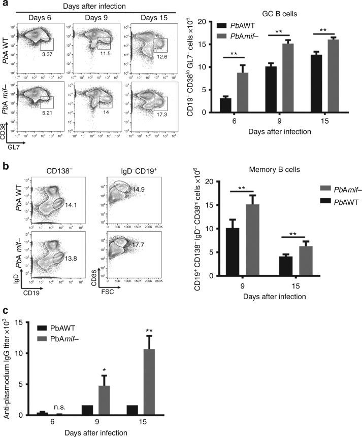
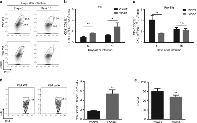
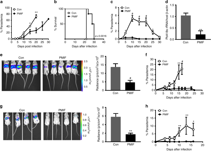
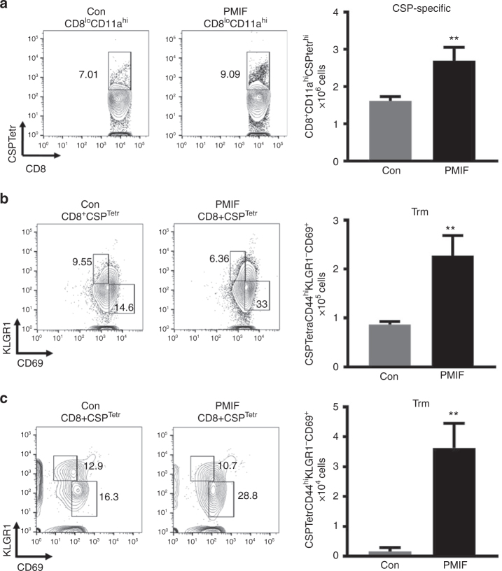
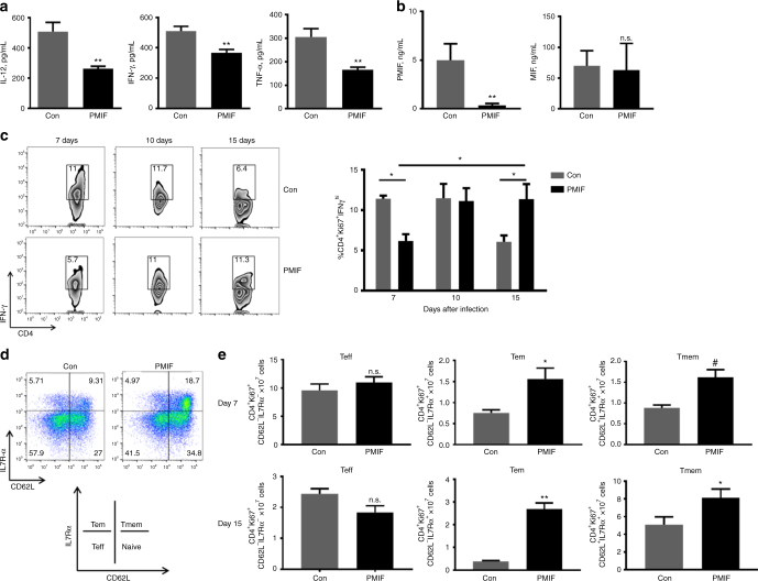
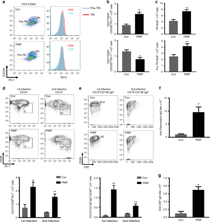
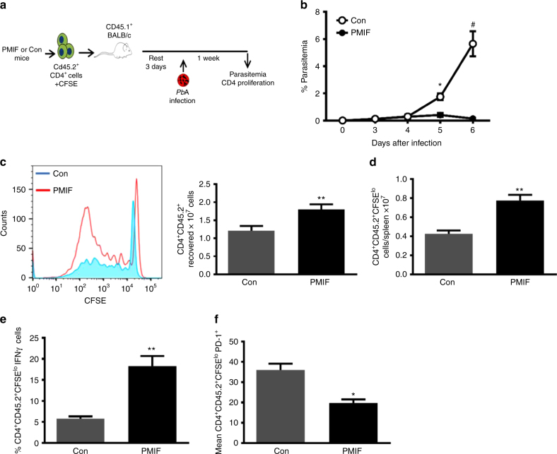
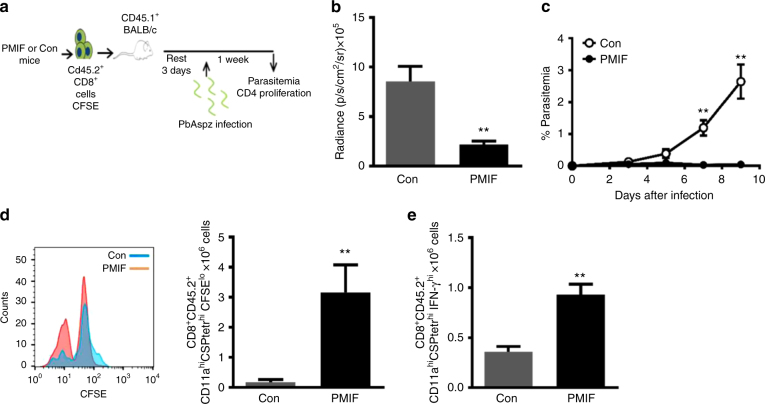
Plasmodium species produce an ortholog of the cytokine macrophage migration inhibitory factor, PMIF, which modulates the host inflammatory response to malaria. Using a novel RNA replicon-based vaccine, we show the impact of PMIF immunoneutralization on the host response and observed improved control of liver and blood-stage Plasmodium infection, and complete protection from re-infection. Vaccination against PMIF delayed blood-stage patency after sporozoite infection, reduced the expression of the Th1-associated inflammatory markers TNF-α, IL-12, and IFN-γ during blood-stage infection, augmented Tfh cell and germinal center responses, increased anti-Plasmodium antibody titers, and enhanced the differentiation of antigen-experienced memory CD4 T cells and liver-resident CD8 T cells. Protection from re-infection was recapitulated by the adoptive transfer of CD8 or CD4 T cells from PMIF RNA immunized hosts. Parasite MIF inhibition may be a useful approach to promote immunity to Plasmodium and potentially other parasite genera that produce MIF orthologous proteins.


Conflict of interest statement

Yale University and Novartis AG have filed a joint patent application describing the potential utility of a pmif encoding RNA replicon. R.B. and A.G. are co-inventors on this application. The remaining authors declare no competing interests.

Figures

Fig. 1
Fig. 1
PMIF impairs germinal center formation. BALB/cJ mice were infected with 106 PbAWT or PbAmif− iRBCs. On day 6, 9, and 15, splenocytes were isolated and the total number of a germinal center (CD19+CD38loGL7+) and b (CD19+CD138IgDCD38hi) memory B cells were determined. Results are from three separate experiments. Bars represent the mean of 12 mice ± SD. **p < 0.05 by Mann–Whitney test. c Anti-Plasmodium antibodies titers from BALB/cJ mice that were infected with PbAWT or PbAmif− iRBCs. On day 6, 9, and 15, sera of infected mice were collected, and the Plasmodium-specific IgG responses measured by ELISA. Results are from three separate experiments. Bars represent the mean of 12 mice ± SD; n.s.: p > 0.05; *p < 0.05; **p < 0.01 by two-way ANOVA
Fig. 2
Fig. 2
PMIF inhibits Tfh cell development. BALB/cJ mice were infected with 106 PbAWT or PbAmif− iRBCs. On days 6 and 15 after infection, splenocytes were isolated and Tfh cells assessed. a–c Representative plots and absolute numbers of Tfh and pre-Tfh cells in the two groups of mice. Results are from three separate experiments. Bars represent the mean of 12 mice ± SD (*p < 0.05, **p < 0.001 by Mann–Whitney test). d Representative plots and absolute number of CD4+CD62LBcl-6hi cells at day 6 after infection, and e expression of transcription factor T-bet in CD4+Bcl-6hi cells. Results are from three separate experiments. Bars represent the mean of 12 mice ± SD; *p < 0.05, **p < 0.001 by Mann–Whitney test
Fig. 3
Fig. 3
PMIF neutralization confers complete protection to re-infection by wild type P. berghei ANKA. a Parasitemia after infection of BALB/cJ mice (106 PbAWT iRBCs) previously immunized with RNA replicons encoding PMIF (black circle) or a control (Con) RNA (white circle); *p < 0.05, **p < 0.01, by two-way ANOVA and error bars denote ±SD. b Kaplan–Meier survival plots for immunized mice following infection with PbAWT (black circle, PMIF and white circle, Con). Data are from two independent experiments with 10–15 animals per group; **p = 0.0016 by log-rank (Mantel Cox) test. c Percentage of iRBCs in BALB/cJ mice previously immunized with RNA encoding PMIF (black circle) or Con RNA (white circle), treated with chloroquine, and re-infected with 106 PbAWT iRBCs; *p < 0.05, #p < 0.0001 by two-way ANOVA and error bars denote ±SD. d Splenic parasite load 6 days after reinfection with iRBCs was measured by quantitative PCR of PbAWT 18S rRNA relative to host β-actin. Results are from two separate experiments. Bars represent the mean of 6 mice ± SD; **p < 0.01 by Mann–Whitney test. e PbAluc liver load and absolute luminescence values in PMIF (black circle) or Con (white circle) RNA replicon immunized mice 48 h after the first infection with 2000 PbAluc sporozoites. f Percentage of iRBCs after the first infection of BALB/cJ mice with 2000 PbAluc sporozoites. Data are from two independent experiments. Bars represent the mean of 10 mice ± SD; **p < 0.01, #p < 0.0001 by Mann–Whitney and two-way ANOVA. g PbA liver load and absolute luminescence values in PMIF (black circle) or Con (white circle) RNA replicon immunized hosts 48 h after the second infection with 2000 PbAluc sporozoites. h Percentage of iRBCs after the second infection of BALB/cJ mice. Data are from two independent experiments. Bars represent the mean of 10 mice ± SD; *p < 0.05, **p < 0.01 by Mann–Whitney test and two-way ANOVA
Fig. 4
Fig. 4
PMIF neutralization enhances the development of Plasmodium liver memory CD8 T cells. BALB/cJ mice immunized with replicons encoding Con RNA or PMIF RNA were challenged with 2000 PbAWT sporozoites by i.v. injection. On day 7 after the first or second infection, liver immune cells were isolated and the percentage and total number of CD8 T cells assessed. a Representative plots and absolute numbers of CSP-specific CD8 T cells (CD8+CD11ahiCSPTetrhi) in the livers of PMIF or Con RNA immunized mice. Results are from two separate experiments. Bars represent the mean of 6 mice ± SD; **p = 0.0022 by Mann–Whitney test. Representative plots and absolute number of CSP-specific tissue resident memory CD8 T cells (Trm: CSPTetrCD44hiKLGR1CD69+) at day 7 after the first (b) and second (c) infection. Results are from two separate experiments. Bars represent the mean of 6 mice ± SD; **p = 0.0022 by Mann–Whitney test
Fig. 5
Fig. 5
PMIF neutralization decreases inflammatory cytokine production and enhances the development of CD4 T cells into effector memory and memory precursors during blood-stage infection. a, b Serum levels of the indicated cytokines were detected by specific ELISA 7 days after injection of 106 PbAWT iRBCs in mice immunized with RNA replicons encoding Con RNA or PMIF RNA. Data are representative of two independent experiments. Bars represent the mean of 6 mice ± SD; *p < 0.05,**p < 0.01 by Mann–Whitney test. c On day 7, 10, and 15 after infection, splenocytes were isolated and stimulated ex vivo with iRBC lysates in the presence of Brefeldin A. Representative dot plots and frequencies of PbAWT responsive CD4 T cells (Ki67+CD4+) expressing IFN-γ in spleens was detected by intracellular staining and analyzed by flow cytometry. Data are representative of two independent experiments. Bars represent the mean of 10 mice ± SD; *p < 0.05 by Mann–Whitney test. d, e Numbers of PbAWT responsive CD4 T cell (Ki67+CD4+) subsets, including T effector (Teff): CD62LIL7Rα, T effector memory (Tem): CD62LIL7Rα+, and T memory (Tmem): CD62L+IL7Rα+ at day 7 and 15 after infection. The contribution of each memory CD4 T cell subset is expressed relative to the total number of PbAWT responsive CD4 T cells. Data are representative of two independent experiments. Bars represent the mean of 10 mice ± SD; n.s.: non-significant, *p < 0.05, **p < 0.001, #p < 0.0001 by Mann–Whitney test
Fig. 6
Fig. 6
PMIF inhibition enhances the development of CD4 Tfh, plasma cells, and anti-Plasmodium antibody responses. BALB/cJ mice immunized with replicons encoding Con RNA or PMIF RNA were infected with 2000 PbAWT sporozoites and splenocytes isolated on day 7 after infection. a, b Representative plots of absolute numbers of Tfh cells (CD4+CD62LCXCR5hiPD-1hi) and pre-Tfh cells (CD4+CD62LCXCR5intPD-1int). c Expression of transcription factor Bcl-6 in Tfh and pre-Tfh cells for both groups of mice during first PbAWT infection. Results are from two separate experiments. Bars represent the mean of 6 mice ± SD; *p < 0.05, **p < 0.001, Ѱp < 0.0001 by Mann–Whitney test. d,e Representative plots and absolute number of germinal center (CD19+CD38loGL7+) and memory B cells (CD19+CD138IgDCD38hi) after the first and second infection. Results are from two separate experiments. Bars represent the mean of 6 mice ± SD; **p < 0.001, #p < 0.0001 by Mann–Whitney test. Serum titers of specific anti-Plasmodium blood-stage (f) and anti-CSP liver-stage antigen (g) IgG from immunized mice analyzed 1 week after the second infection with PbAWT sporozoites. Data shown are from three and two independent experiments, respectively. Bars represent the mean of 10 mice ± SD; #p < 0.0001 by Mann–Whitney test
Fig. 7
Fig. 7
Adoptively transferred CD4 T cells from PMIF-immunized mice confer protection to challenge by iRBCs. a BALB/cJ mice immunized with replicons encoding Con RNA or PMIF RNA were infected with 106 PbAWT iRBCs and treated with chloroquine on days 7–12. Four weeks later, the mice were reinfected with 106 PbAWT iRBCs and splenocytes isolated 7 days after infection, incubated with chloroquine to eliminate blood-stage parasites, and labeled with CFSE. Purified CD4+CD45.2+ T cells (2 × 107) then were transferred into naïve congenic CD45.1 BALB/cJ hosts and the mice infected 3 days later with 106 PbAWT iRBCs. b Frequency of iRBCs in mice adoptively transferred with CD4 T cells from Con (white circle) or PMIF (black circle) RNA immunized mice. Results are from two separate experiments. Bars represent the mean of 6 mice ± SD; *p < 0.05, #p < 0.001 by two-way ANOVA. c Representative CFSE dilution histogram of adoptively transferred CD4+ T cells (CD45.2) from donors immunized with Con or PMIF RNA and enumeration of recovered CD45.2 CD4+ T cells, and d proliferative response of transferred CD4 T cells into CD45.1 recipients 7 days after infection. e Percentage of proliferating CD45.2+ CD4+ T cells (CFSElo) producing IFN-γ after stimulation ex vivo with iRBC lysates in the presence of Brefeldin A. f Mean fluorescence intensity of PD-1 in PbAWT responsive CD45.2+ CD4+ T cells (CFSElo) from Con or PMIF RNA immunized donors. Results are from two separate experiments. Bars represent the mean of 8 mice ± SD; *p < 0.05, **p < 0.01 by two-tailed Mann–Whitney test
Fig. 8
Fig. 8
Adoptively transferred liver CD8 T cells from PMIF-immunized mice confer protection to homologous sporozoite challenge. a BALB/cJ mice immunized with replicons encoding Con RNA or PMIF RNA were infected with 2000 PbAWT sporozoites and cured by 6 days of chloroquine treatment (days 7–12). Four weeks later, the mice were reinfected with 2000 PbAWT sporozoites and T cells from liver isolated 7 days after infection, incubated with chloroquine to eliminate residual blood-stage parasites, and labeled with CFSE. Purified CD45.2+ CD8 T cells (2 × 107) then were transferred into naïve congenic CD45.1 BALB/cJ hosts and the mice infected 3 days later with 2000 PbAWT sporozoites. b Luminescence values of infected mice and c parasitemia in mice adoptively transferred with liver CD8 T cells from Con RNA (white circle) or PMIF RNA (black circle) immunized mice. Results are from two separate experiments. Bars represent the mean of 6 mice ± SD **p < 0.01 by two-way ANOVA. d Representative CFSE dilution histogram of adoptively transferred (CD45.2) CD8 T cells from Con RNA or PMIF RNA immunized donors and enumeration of recovered CD45.2 CD8 T cells. e Number of proliferating CD45.2 CD8 T cells (CFSElo) producing IFN-γ after stimulation ex vivo with CSP peptide in the presence of Brefeldin A. Results are from two separate experiments. Bars represent the mean of 6 mice ± SD;**p < 0.01 by two-tailed Mann–Whitney test, error bars denote ±SD

Comment in

  • Novel malaria vaccines.
    Laurens MB. Laurens MB. Hum Vaccin Immunother. 2021 Nov 2;17(11):4549-4552. doi: 10.1080/21645515.2021.1947762. Epub 2021 Aug 4. Hum Vaccin Immunother. 2021. PMID: 34347570 Free PMC article.

References

    1. World Health Organization. World Malaria Report 2014 (World Health Organization, 2014).
    1. Langhorne J, Ndungu FM, Sponaas AM, Marsh K. Immunity to malaria: more questions than answers. Nat. Immunol. 2008;9:725–732. doi: 10.1038/ni.f.205. - DOI - PubMed
    1. Tran TM, et al. An intensive longitudinal cohort study of Malian children and adults reveals no evidence of acquired immunity to Plasmodium falciparum infection. Clin. Infect. Dis. 2013;57:40–47. doi: 10.1093/cid/cit174. - DOI - PMC - PubMed
    1. Riley EM, Stewart VA. Immune mechanisms in malaria: new insights in vaccine development. Nat. Med. 2013;19:168–178. doi: 10.1038/nm.3083. - DOI - PubMed
    1. Wykes, M. N., Horne-Debets, J. M., Leow, C.-Y. & Karunarathne, D. Malaria drives T cells to exhaustion. Front. Microbiol.5, 249 (2014). - PMC - PubMed

Publication types

MeSH terms