. 2018 Jul 13;9(1):2704.
doi: 10.1038/s41467-018-05039-1.

HHEX is a transcriptional regulator of the VEGFC/FLT4/PROX1 signaling axis during vascular development

Affiliations

HHEX is a transcriptional regulator of the VEGFC/FLT4/PROX1 signaling axis during vascular development

Sébastien Gauvrit et al. Nat Commun. .

Abstract

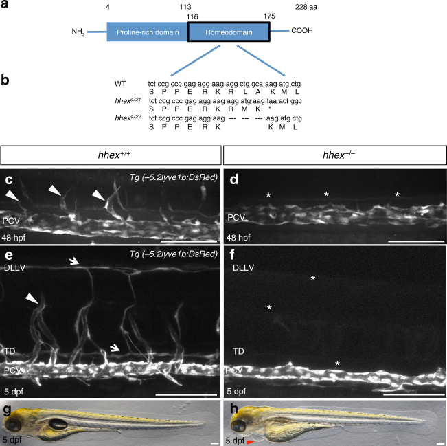
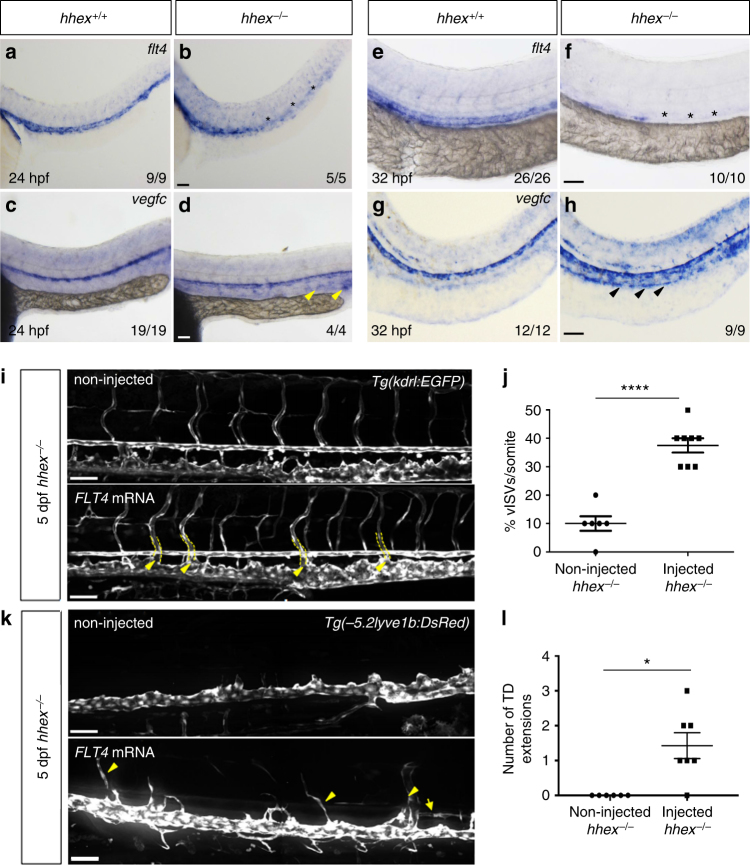
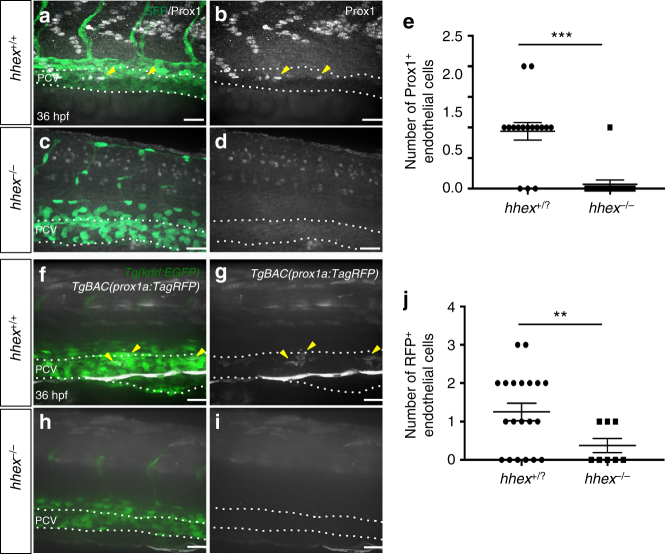
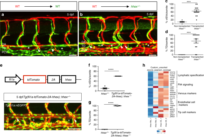
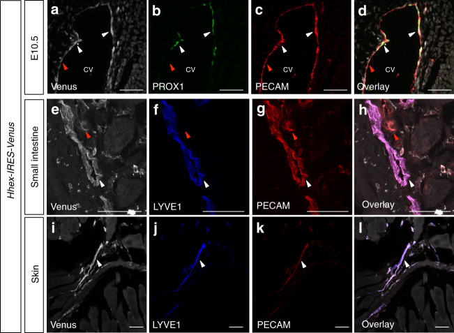
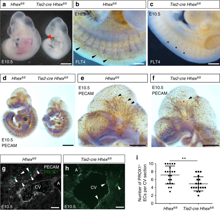
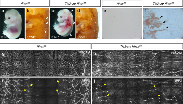
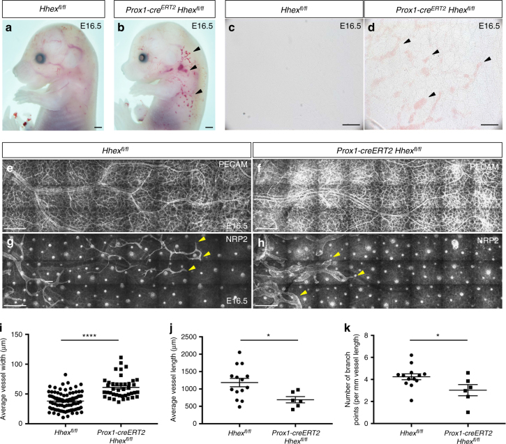
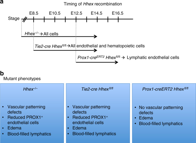
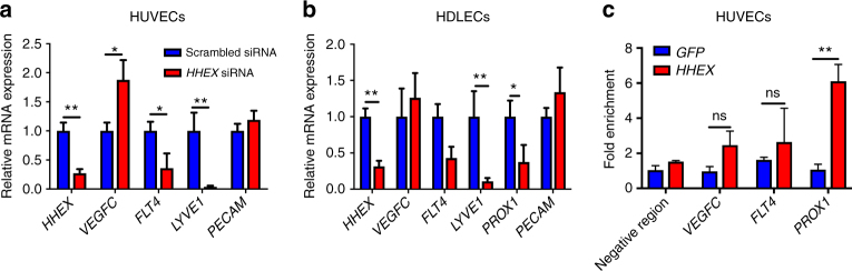
Formation of the lymphatic system requires the coordinated expression of several key regulators: vascular endothelial growth factor C (VEGFC), its receptor FLT4, and a key transcriptional effector, PROX1. Yet, how expression of these signaling components is regulated remains poorly understood. Here, using a combination of genetic and molecular approaches, we identify the transcription factor hematopoietically expressed homeobox (HHEX) as an upstream regulator of VEGFC, FLT4, and PROX1 during angiogenic sprouting and lymphatic formation in vertebrates. By analyzing zebrafish mutants, we found that hhex is necessary for sprouting angiogenesis from the posterior cardinal vein, a process required for lymphangiogenesis. Furthermore, studies of mammalian HHEX using tissue-specific genetic deletions in mouse and knockdowns in cultured human endothelial cells reveal its highly conserved function during vascular and lymphatic development. Our findings that HHEX is essential for the regulation of the VEGFC/FLT4/PROX1 axis provide insights into the molecular regulation of lymphangiogenesis.


Conflict of interest statement

The authors declare no competing interests.

Figures

Fig. 1
Fig. 1
Zebrafish hhex mutants lack sprouting angiogenesis from the posterior cardinal vein. a Schematic representation of Hhex. Hhex, 228 amino acids (aa) long, is composed of a proline-rich domain (4–113 aa) and a homeodomain (116–175 aa). b Alignment of partial Hhex homeodomain sequence in wild-type (WT), and two mutant alleles, hhexs721 and hhexs722. The hhexs721 allele contains a 10 bp insertion leading to a premature stop codon within the homeodomain coding region, whereas the hhexs722 allele lacks amino acids R149 to A151. c, d Trunk vasculature of Tg(−5.2lyve1b:DsRed); hhex+/+ and hhex−/− embryos at 48 hpf. hhex mutant trunks exhibit a defect in sprouting angiogenesis from the posterior cardinal vein (PCV) (arrowheads point to tip cells sprouting from the PCV; asterisks indicate lack of tip cells sprouting from the PCV). e, f Trunk vasculature of Tg(−5.2lyve1b:DsRed); hhex+/+ and hhex−/− larvae at 5 dpf. hhex mutant trunks exhibit a defect in the formation of the venous intersegmental vessels (vISVs), the thoracic duct (TD) lymphatic vessel, and the dorsal longitudinal lymphatic vessel (DLLV) (arrowhead points to a vISV; arrows point to the ventrally positioned TD and dorsally positioned DLLV; asterisks indicate lack of these structures). g, h Brightfield lateral views of hhex+/+ and hhex−/− larvae at 5 dpf. Mutant larvae exhibit pericardial edema (arrowhead). Scale bars: 100 μm
Fig. 2
Fig. 2
The Vegfc/Flt4 pathway is affected in zebrafish hhex mutants. ad Whole-mount in situ hybridization showing flt4 (a, b) and vegfc (c, d) expression in 24 hpf hhex+/+ and hhex−/− embryos. At 24 hpf, hhex mutants exhibit decreased flt4 expression (asterisks), whereas vegfc expression in the PCV appears to be slightly increased (arrowheads). eh Whole-mount in situ hybridization showing flt4 (e, f) and vegfc (g, h) expression at 32 hpf in hhex+/+ and hhex−/− embryos. At 32 hpf, hhex mutants exhibit a strong decrease in flt4 expression (asterisks), whereas vegfc expression is clearly increased in the PCV (arrowheads). x/y: number of embryos showing representative phenotype (x), number of embryos examined (y). i Trunk vasculature of 5 dpf Tg(kdrl:EGFP); hhex−/− injected, or not, with full-length human FLT4 mRNA. hhex mutants exhibit partial rescue of their vISVs at 5 dpf (arrowheads point to vISVs). j Quantification of vISVs across 10 somites in 5 dpf non-injected hhex−/− (n = 6) and FLT4 mRNA-injected hhex−/− (n = 8). k Trunk lymphatic vasculature of 5 dpf Tg(−5.2lyve1b:DsRed); hhex−/− injected, or not, with full-length human FLT4 mRNA. hhex mutants exhibit partial rescue of their TD at 5 dpf (arrowheads point to vISVs and arrow points to TD). l Quantification of TD extensions across 10 somites in 5 dpf non-injected hhex−/− (n = 6) and FLT4 mRNA-injected hhex−/− (n = 7). Values represent means ± s.e.m. ****P ≤ 0.0001 and *P ≤ 0.05 by t-test. Scale bars: 50 μm
Fig. 3
Fig. 3
Zebrafish hhex mutants lack lymphatic precursors. ad Whole-mount views of 36 hpf hhex+/+ and hhex−/− embryos immunostained for Prox1 (arrowheads point to Prox1+ endothelial cells in the PCV). e Quantification of the number of Prox1+ endothelial cells across three somites (from two field of view per embryo) in hhex+/? (n = 8) and hhex−/− (n = 6). fi Whole-mount views of 36 hpf Tg(prox1a:TagRFP); hhex+/+ and hhex−/− embryos. hhex mutants exhibit a strong decrease in the number of RFP+ cells in the PCV compared to hhex+/+ (arrowheads point to RFP+ endothelial cells in the PCV). j Quantification of the number of RFP+ endothelial cells across three somites (from two field of view per embryo) in hhex+/? (n = 10) and hhex−/− (n = 4). Values represent means ± s.e.m. ***P ≤ 0.001, and **P ≤ 0.01 by t-test. Scale bars: 50 μm
Fig. 4
Fig. 4
Hhex is required cell-autonomously in endothelial cells to promote venous and lymphatic sprouting in zebrafish. a, b Transplantation of Tg(fli1ep:DsRedEx) donor cells into TgBAC(etv2:EGFP) hosts derived from hhex+/− incrosses. Wild-type endothelial cells contribute to arteries, veins, and lymphatics in wild-type sibling (a) and mutant (b) hosts at 5 dpf (arrowheads point to vISVs; asterisks indicate TD). c, d Quantification of vISVs (c) and TD extensions (d) across four somites in hhex−/− larvae with transplanted wild-type cells (n = 13) vs. hhex−/− larvae without transplanted wild-type cells (n = 6) at 5 dpf. Wild-type endothelial cells can partially rescue both vISV and TD formation in hhex−/−. e hhex endothelial overexpression strategy using the fli1a promoter partially rescues the hhex−/− vascular phenotype (arrowheads point to vISVs; asterisks indicate TD). f, g Quantification of vISVs (f) and TD (g) extensions across four somites in hhex−/− (n = 6) and Tg(fli1a:tdTomato-2A-hhex); hhex−/− (n = 6). h RNA sequencing of 48 hpf FACS-sorted hhex−/− endothelial cells and hhex-overexpressing endothelial cells. Heat map comparisons between these datasets identify Hhex as a regulator of genes implicated in lymphatic specification (prox1a, prox1b, mafba, sox18, nr2f2) and Flt4 signaling (nrp2a, nrp2b, flt4, vegfc) while endothelial cell markers are modulated only by endothelial-specific hhex overexpression. Values represent means ± s.e.m. ****P ≤ 0.0001 and ***P ≤ 0.001 by t-test. Scale bars: 50 μm
Fig. 5
Fig. 5
Hhex-IRES-Venus is expressed by vascular endothelial cells and lymphatic endothelial cells during mouse development and adulthood. ad Maximum intensity projections of confocal images of an E10.5 Hhex-IRES-Venus embryo in the CV region after Venus (white), PROX1 (green), and PECAM (red) immunostaining. el Maximum intensity projections of confocal images of an Hhex-IRES-Venus adult mouse small intestine (eh) and skin (il) after Venus (white), LYVE1 (blue) and PECAM (red) immunostaining. Hhex-IRES-Venus expression is observed in blood endothelial cells (red arrowheads) as well as LECs (white arrowheads). Scale bars: 50 μm
Fig. 6
Fig. 6
Tie2-cre Hhexfl/fl mouse embryos exhibit minor vascular defects and a reduced number of PROX1+ endothelial cells at E10.5. a Whole-mount view of Hhexfl/fl and Tie2-cre Hhexfl/fl embryos at E10.5. Hhex mutants exhibit pericardial edema (red arrowhead) as well as developmental delay. b, c Whole-mount views of Hhexfl/fl and Tie2-cre Hhexfl/fl embryos at E10.5 after FLT4 immunostaining. Hhex mutants exhibit a strong decrease in FLT4 expression in the intersomitic vessels (arrowheads point to intersomitic vessels; asterisks indicate reduction of FLT4 expression in the same structures). df Whole-mount view of Hhexfl/fl and Tie2-cre Hhexfl/fl embryos at E10.5 after PECAM immunostaining. Hhex mutants exhibit minor vascular defects in the head (arrowheads point to blood vessels in Hhexfl/fl and Tie2-cre Hhexfl/fl; asterisk indicates lack of vessels in the same structures in Tie2-cre Hhexfl/fl). g, h Maximum intensity projections of confocal images from transverse cryosections of Hhexfl/fl and Tie2-cre Hhexfl/fl E10.5 embryos after immunostaining for PECAM (white) and PROX1 (green) in the cardinal vein (CV) and aorta (A) region. Hhex mutants exhibit fewer PROX1+/PECAM+ endothelial cells in the region of the CV (arrowheads point to PROX1+/PECAM+ endothelial cells). i Quantification of the number of PROX1+ endothelial cells in the CV region in Hhexfl/fl (n = 2) and Tie2-cre Hhexfl/fl (n = 2) embryos. Values represent means ± s.e.m. **P ≤ 0.01 by t-test. Scale bars: 1 mm (a, d), 200 μm (b, c), 500 μm (e, f), and 20 μm (g, h)
Fig. 7
Fig. 7
Tie2-cre Hhexfl/fl mouse embryos exhibit strong vascular and lymphatic defects at E14.5. ac Morphology of Hhexfl/fl and Tie2-cre Hhexfl/fl mouse embryos at E14.5. Hhex inactivation in Tie2-expressing cells leads to lymphatic defects including edema and blood-filled lymphatics (arrowheads). bd Whole-mount views of Hhexfl/fl and Tie2-cre Hhexfl/fl embryos at E14.5 after LYVE1 immunostaining. Hhex mutants exhibit defects in lymphatic patterning with a reduced lymphatic vasculature in the skin (arrowheads indicate LYVE1+ lymphatic vessels, asterisks indicate lack of these vessels in the same area in Tie2-cre Hhexfl/fl). e, f Brightfield pictures of Hhexfl/fl and Tie2-cre Hhexfl/fl skin at E14.5. Hhex mutants exhibit blood-filled lymphatics (arrowheads). g, h Whole-mount views of Hhexfl/fl and Tie2-cre Hhexfl/fl skin at E14.5 after PECAM immunostaining. Hhex mutants exhibit strong vascular defects at E14.5. i, j Whole-mount views of Hhexfl/fl and Tie2-cre Hhexfl/fl skin at E14.5 after NRP2 immunostaining. Hhex mutants exhibit strong lymphatic defects at E14.5 (arrows point to lymphatic vessels; arrowheads point to blood-filled lymphatic vessels with decreased intensity of NRP2 immunostaining). Scale bars: 2 mm (a, c), 1 mm (b, d), 200 μm (e, f), and 250 μm (gj)
Fig. 8
Fig. 8
Prox1-creERT2 Hhexfl/fl mouse embryos exhibit lymphatic defects at E16.5. a, b Whole-mount views of Hhexfl/fl and Prox1-creERT2 Hhexfl/fl embryos at E16.5 after tamoxifen injection at E10.5, 11.5, and 12.5. Hhex deletion in Prox1-expressing cells leads to lymphatic defects including edema and blood-filled lymphatics (arrowheads). c, d Brightfield pictures of Hhexfl/fl and Prox1-creERT2 Hhexfl/fl skin at E16.5. Hhex mutants exhibit blood-filled lymphatics (arrowheads). eh Whole-mount views of Hhexfl/fl and Prox1-creERT2 Hhexfl/fl skin at E16.5 after PECAM (ef) and NRP2 (g, h) immunostaining. Hhex mutants do not exhibit vascular defects but clear lymphatic defects (arrowheads point to lymphatic vessels). ik Quantification of the lymphatic network including average vessel width (i), average vessel length (j), and number of branch points (per mm vessel length) (k) in Hhexfl/fl (n = 3) and Prox1-creERT2 Hhexfl/fl (n = 3) skin at E16.5. Values represent means ± s.e.m. ****P ≤ 0.0001 and *P ≤ 0.05 by t-test. Scale bars: 1 mm (a, b), 200 μm (c, d), and 250 μm (eh)
Fig. 9
Fig. 9
Summary of the effects of global and conditional Hhex deletion during vascular development in mouse. a Stages when Hhex recombination occurs in Hhex−/, Tie2-cre Hhexfl/fl, and Prox1-creERT2 Hhexfl/fl. b Consequences of Hhex deletion in Hhex-/-, Tie2-cre Hhexfl/fl, and Prox1-creERT2 Hhexfl/f are summarized in the blue boxes
Fig. 10
Fig. 10
HHEX regulates the VEGFC/FLT4/PROX1 pathway in human endothelial cells and binds the PROX1 promoter. a, b qPCR analysis of HHEX, VEGFC, FLT4, LYVE1, PROX1, and PECAM expression in HUVECs (a) and HDLECs (b) treated with HHEX siRNA compared to control cells treated with a scrambled siRNA. Knockdown of HHEX led to downregulation of FLT4, PROX1, and LYVE1 and upregulation of VEGFC expression. c Chromatin immunoprecipitation (ChIP) experiment using Myc-HHEX adenovirus in HUVECs. qPCR using primers for specific FLT4, VEGFC, and PROX1 regions was carried out and enrichment relative to the percentage of input in the IP sample at PRM2 (control region) is shown. HHEX does not appear to bind the FLT4 promoter or VEGFC 3′ region but strongly binds the PROX1 promoter. Values represent means ± s.e.m. **P ≤ 0.01; *P ≤ 0.05; ns, not significant by Mann-Whitney (a, b) or Bonferroni post hoc (c)

References

    1. Karkkainen MJ, et al. Vascular endothelial growth factor C is required for sprouting of the first lymphatic vessels from embryonic veins. Nat. Immunol. 2004;5:74–80. doi: 10.1038/ni1013. - DOI - PubMed
    1. Zhang L, et al. VEGFR-3 ligand-binding and kinase activity are required for lymphangiogenesis but not for angiogenesis. Cell Res. 2010;20:1319–1331. doi: 10.1038/cr.2010.116. - DOI - PubMed
    1. Wigle JT, Oliver G. Prox1 function is required for the development of the murine lymphatic system. Cell. 1999;98:769–778. doi: 10.1016/S0092-8674(00)81511-1. - DOI - PubMed
    1. Newman CS, Chia F, Krieg PA. The XHex homeobox gene is expressed during development of the vascular endothelium: overexpression leads to an increase in vascular endothelial cell number. Mech. Dev. 1997;66:83–93. doi: 10.1016/S0925-4773(97)00092-0. - DOI - PubMed
    1. Liao W, Ho CY, Yan YL, Postlethwait J, Stainier DY. Hhex and scl function in parallel to regulate early endothelial and blood differentiation in zebrafish. Development. 2000;127:4303–4313. - PubMed

Publication types

MeSH terms

Substances