. 2018 Jul 13;9(1):2716.
doi: 10.1038/s41467-018-05288-0.

Nonstimulatory peptide-MHC enhances human T-cell antigen-specific responses by amplifying proximal TCR signaling

Affiliations

Nonstimulatory peptide-MHC enhances human T-cell antigen-specific responses by amplifying proximal TCR signaling

Xiang Zhao et al. Nat Commun. .

Abstract

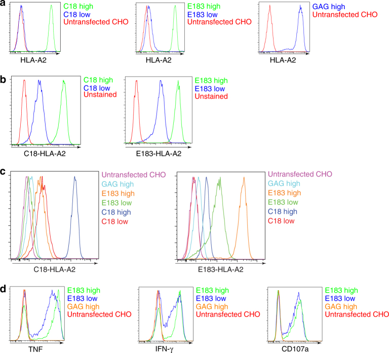
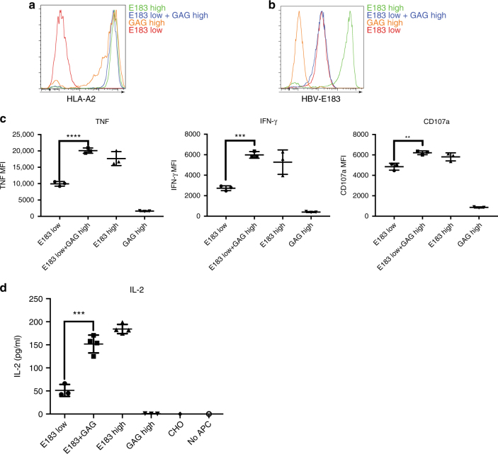
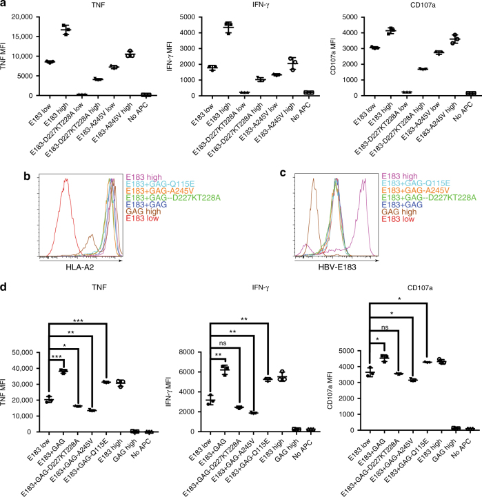
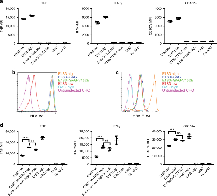
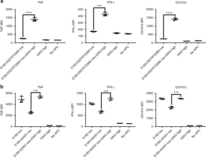
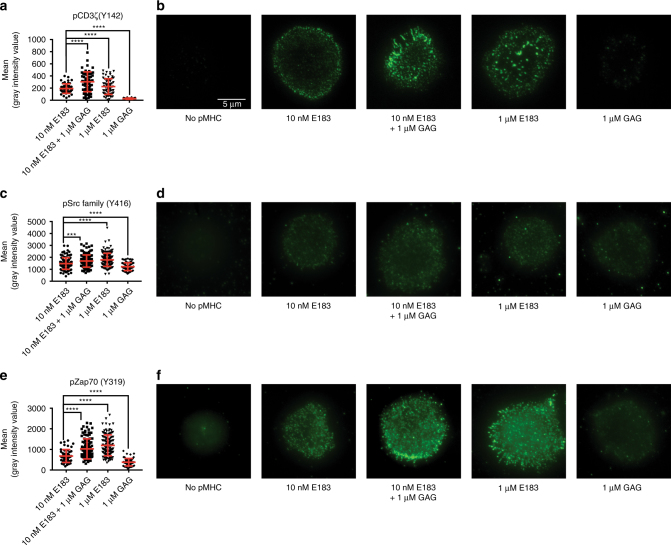
Foreign antigens are presented by antigen-presenting cells in the presence of abundant endogenous peptides that are nonstimulatory to the T cell. In mouse T cells, endogenous, nonstimulatory peptides have been shown to enhance responses to specific peptide antigens, a phenomenon termed coagonism. However, whether coagonism also occurs in human T cells is unclear, and the molecular mechanism of coagonism is still under debate since CD4 and CD8 coagonism requires different interactions. Here we show that the nonstimulatory, HIV-derived peptide GAG enhances a specific human cytotoxic T lymphocyte response to HBV-derived epitopes presented by HLA-A*02:01. Coagonism in human T cells requires the CD8 coreceptor, but not T-cell receptor (TCR) binding to the nonstimulatory peptide-MHC. Coagonists enhance the phosphorylation and recruitment of several molecules involved in the TCR-proximal signaling pathway, suggesting that coagonists promote T-cell responses to antigenic pMHC by amplifying TCR-proximal signaling.


Conflict of interest statement

The authors declare no competing interests.

Figures

Fig. 1
Fig. 1
Inducible expression of human single-chain (sc)-MHCI in T-REx CHO cells and their immunogenicity to human CTL. a anti-HLA-A2 staining of T-REx CHO clones expressing low amount or high amounts of (doxycycline-induced) human single-chain (sc)-MHCI. b TCR-like antibody anti-C18-HLA-A2 or anti-E183-HLA-A2 staining of T-REx CHO clones expressing low or high concentrations of C18/E183-HLA-A2 sc pMHC. c TCR-like antibody anti-C18-HLA-A2 or anti-E183-HLA-A2 staining of T-REx CHO clones expressing low or high C18/E183-HLA-A2 sc pMHC, high concentration of GAG-HLA-A2 sc pMHC, and unstransfected T-REx CHO cells. d Human E183 epitope-specific CTL were cultured with the indicated T-REx CHO cells for 3 h and cytokine expression (TNF and IFN-γ) and upregulation of the degranulation marker CD107a were assessed by flow cytometry. Data are representative of five independent experiments
Fig. 2
Fig. 2
Nonstimulatory pMHCI can enhance human CTL response to antigen. a Anti-HLA-A2 staining of T-REx CHO clones used in the coagonism experiments. b Measurement of the amount of the antigen, E183-HLA-A2 in T-REx CHO clones by staining with TCR-like antibody anti-E183-HLA-A2. c Human E183 CTL were cultured with the indicated T-REx CHO clones panel for 3 h and cytokine expression (TNF and IFN-γ) and upregulation of the degranulation marker CD107a were assessed by flow cytometry. d Human E183 CTL were cultured with the indicated T-REx CHO clones panel for 24 h, and the amount of IL-2 in the supernatant was assessed by ELISA. Statistical significance was determined by using unpaired two-sided Student’s t test and shown as mean ± s.d. (*p < 0.05, **p < 0.01, ***p < 0.001). Data are representative of five independent experiments. MFI mean fluorescence level
Fig. 3
Fig. 3
CD8 binding to nonstimulatory pMHCI is required for coagonism in human T cells. a D227K, T228A, or A245V mutations were introduced in the antigen sc E183-HLA-A2 (sc E183-HLA-A2-CD8mut). Doxycycline-inducible sc E183-HLA-A2-CD8mut constructs were transfected into T-REx CHO cells. Single cell clones were used to stimulate human E183 CTL for 3 h. Cytokine production (TNF and IFN-γ) and upregulation of the degranulation marker CD107a in E183 CTL were assessed by flow cytometry. b D227K, T228A, A245V, or Q115E mutations were introduced in the nonstimulatory sc GAG-HLA-A2 (sc GAG-HLA-A2-CD8mut). sc GAG-HLA-A2-CD8mut constructs were transfected into the T-REx CHO clone that could simultaneously express low concentration of the antigen sc E183-HLA-A2 and single cell clones were sorted. Anti-HLA-A2 staining (b) or anti-E183-HLA-A2 staining using TCR-like antibody (c) was performed on the indicated T-REx CHO clones. d Human E183 CTL were cultured with the indicated T-REx CHO cells for 3 h and cytokine production (TNF and IFN-γ) and upregulation of the degranulation marker CD107a were assessed by flow cytometry. Statistical significance was determined by using unpaired two-sided Student’s t test and shown as mean ± s.d. (ns nonsignificant, *p < 0.05, **p < 0.01, ***p < 0.001, ****p < 0.0001). Data are representative of three independent experiments. MFI mean fluorescence level
Fig. 4
Fig. 4
TCR binding to nonstimulatory pMHCI is not required for coagonism in human T cells. a V152E mutation was introduced into the antigen sc E183-HLA-A2 which was then transfected into T-REx CHO cells and single cell clones were sorted. Indicated T-REx CHO clones panel was cocultured with human E183 CTL for 3 h. Cytokine production (TNF and IFN-γ) and upregulation of the degranulation marker CD107a were assessed by antibody staining and flow cytometry. b V152E mutation was introduced into nonstimulatory sc GAG-HLA-A2 which was then transfected into the T-REx CHO clone that could simultaneously express low concentration of the antigen sc E183-HLA-A2. Anti-HLA-A2 (b) or anti-E183-HLA-A2 (c) staining was performed on the indicated T-REx CHO cells and analyzed by flow cytometry. d Human E183 CTL were cocultured with the indicated T-REx CHO clones panel for 3 h. Cytokine production (TNF and IFN-γ) and upregulation of the degranulation marker CD107a were assessed by antibody staining and flow cytometry. Statistical significance was determined by using unpaired two-sided Student’s t test and shown as mean ± s.d. (ns nonsignificant, *p < 0.05, **p < 0.01, ***p < 0.001, ****p < 0.0001). Data are representative of three independent experiments. MFI mean fluorescence level
Fig. 5
Fig. 5
Nonstimulatory pMHC can enhance T-cell activation when CD8 binding to antigenic pMHCI is abrogated. sc GAG-HLA-A2 constructs were transfected into the T-REx CHO clone that could express low amount sc E183-HLA-A2 bearing D227K,T228A (a) or A245V (b) mutation. Single cell clones were sorted. Indicated T-REx CHO clones were co-cultured with human E183 CTL for 3 h. Cytokine production (TNF and IFN-γ) and upregulation of the degranulation marker CD107a were assessed by antibody staining and flow cytometry. Statistical significance was determined by using unpaired two-sided Student’s t test and shown as mean ± s.d. (ns nonsignificant, *p < 0.05, **p < 0.01, ***p < 0.001, ****p < 0.0001). Data are representative of three independent experiments. MFI mean fluorescence level
Fig. 6
Fig. 6
CD3 and CD8 are not strongly downregulated in coagonism. Anti-CD3 (a) and anti-CD8 (b) staining were performed in human CTL activation experiments stimulated by the indicated T-REx CHO clones. CD3 and CD8 amount were then assessed by flow cytometry. Data are representative of three independent experiments. MFI mean fluorescence level
Fig. 7
Fig. 7
Coagonist pMHC enhance human T-cell response to antigens by increasing phosphorylation of molecules involved in proximal TCR signaling pathway. Human E183 CTL were stimulated by T-REx CHO cells panel for 5 or 30 min, lysed and the lysate was separated on NuPAGE Bis-Tris Gel. The filter was blotted with antibodies against pCD3ζ (a), pZap70 (b), pLAT (c), pPLC-γ1 (d), p-Erk1/2 (e), and pSHP-1 (f). The data are representative of three independent experiments. Uncropped western blotting images are presented in Supplementary Fig. 8
Fig. 8
Fig. 8
Coagonist pMHC enhance the recruitment of pCD3ζ, pLck, and pZap70 into the immunological synapse. Different concentrations of E183-A2 monomer or GAG-A2 monomer were added to the lipid bilayers in a glass chamber slide. Totally, 105 E183 CTL were added into each well of the chamber and stimulated for 5 min. The CTL were fixed, permeabilized and stained with different antibodies to check the recruitment of pCD3ζ (a, b), pLck (c, d), and pZap70 (e, f) into the immunological synapse by TIRF microscopy. Anti-pSrc (Y416) antibody was used to detect active Lck (p-Y394). Statistical significance was determined by using unpaired two-sided Student’s t test and shown as mean ± s.d. (ns nonsignificant, *p < 0.05, **p < 0.01, ***p < 0.001, ****p < 0.0001). Data are representative of three independent experiments
Fig. 9
Fig. 9
Analog amplifier model for the mechanism of coagonism. Nonstimulatory pMHC molecules recruit CD8-Lck into the immunological synapse. Preconcentrated Lck further phosphorylate the ITAMs of TCR-CD3ζ which are engaged with specific antigenic pMHC. Enhanced pCD3ζ provide more docking sites for Zap70 to be phosphorylated. Enhanced pZap70 signal amplifies the downstream TCR signaling pathway including the phosphorylation of Erk. pErk translocates into the nucleus and enhances the effector functional readouts of T-cell activation

Similar articles

Cited by

References

    1. Irvine DJ, Purbhoo MA, Krogsgaard M, Davis MM. Direct observation of ligand recognition by T cells. Nature. 2002;419:845–849. doi: 10.1038/nature01076. - DOI - PubMed
    1. Gascoigne NRJ. Do T cells need endogenous peptides for activation? Nat. Rev. Immunol. 2008;8:895–900. doi: 10.1038/nri2431. - DOI - PubMed
    1. Krogsgaard M, et al. Agonist/endogenous peptide–MHC heterodimers drive T-cell activation and sensitivity. Nature. 2005;434:238–243. doi: 10.1038/nature03391. - DOI - PubMed
    1. Yachi PP, Ampudia J, Gascoigne NRJ, Zal T. Nonstimulatory peptides contribute to antigen-induced CD8 T-cell receptor interaction at the immunological synapse. Nat. Immunol. 2005;6:785–792. doi: 10.1038/ni1220. - DOI - PMC - PubMed
    1. Yachi PP, Lotz C, Ampudia J, Gascoigne NRJ. T-cell activation enhancement by endogenous pMHC acts for both weak and strong agonists but varies with differentiation state. J. Exp. Med. 2007;204:2747–2757. doi: 10.1084/jem.20062610. - DOI - PMC - PubMed

Publication types

MeSH terms