Review
. 2018 Nov;592(21):3586-3605.
doi: 10.1002/1873-3468.13192. Epub 2018 Jul 28.

Molecular mechanisms of force production in clathrin-mediated endocytosis

Affiliations
Review

Molecular mechanisms of force production in clathrin-mediated endocytosis

Michael M Lacy et al. FEBS Lett. 2018 Nov.

Abstract

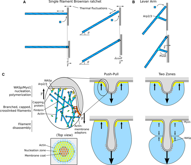
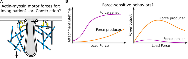
During clathrin-mediated endocytosis (CME), a flat patch of membrane is invaginated and pinched off to release a vesicle into the cytoplasm. In yeast CME, over 60 proteins-including a dynamic actin meshwork-self-assemble to deform the plasma membrane. Several models have been proposed for how actin and other molecules produce the forces necessary to overcome the mechanical barriers of membrane tension and turgor pressure, but the precise mechanisms and a full picture of their interplay are still not clear. In this review, we discuss the evidence for these force production models from a quantitative perspective and propose future directions for experimental and theoretical work that could clarify their various contributions.

Keywords: actin; clathrin; endocytosis; membrane; membrane remodeling; yeast.


Figures

Fig. 1.
Fig. 1.
Overview of proteins and forces in clathrin-mediated endocytosis in yeast. (A) Stages of CME membrane deformation, and spatial organization and timing of various protein modules. Membrane shapes, actin filaments, and vesicle are drawn to scale, reflecting quantitative microscopy data from yeast. Myosin-I and WASp localizations are represented by dashed lines when the reported localizations in S. cerevisiae and S. pombe differ. (B) Forces opposing CME. Turgor pressure, membrane bending and membrane tension pose significant energy barriers that must be overcome to generate a clathrin-coated pit and vesicle. Note that turgor pressure is applied isotropically to all membrane surfaces, favoring collapse of the pit and tubule, and membrane scission passes through a high-energy intermediate. Arrows are drawn to indicate the direction and order of magnitude of forces opposing CME.
Fig. 2.
Fig. 2.
Actin force production by polymerization. (A) Brownian ratchet model for force production from polymerization of a single filament. Left: A single filament polymerizing against a barrier or object exerts force related to the single polymerization step distance d. Right: A filament at an angle exerts force related to the step distance dcosh. If the filament is maintained at an angle (e.g., as one branch in a meshwork), the stall force is higher but the velocity of the barrier object is lower compared with the perpendicular filament. (B) Actin polymerization force can be distributed through pivot points. Polymerizing filaments exert force not only at their barbed end but may also generate torque with branched or crosslinked filaments or membrane-bound proteins acting as a lever arm. (C) Schematic of the dendritic nucleation model for the endocytic actin meshwork. Left inset: Force production can be achieved by WASp/Myo1 nucleation at the membrane surface, actin filament branching and polymerization, capping and crosslinking, and attachment to the invaginating CCP tip to transmit force from the growing meshwork. Right: The Push-Pull model proposes an actin meshwork nucleated at the base membrane pushing toward the cytoplasm and attachment to the CCP tip pulling the membrane. Far right: The two-zone model proposes that, as the CCP elongates, two distinct zones of nucleation (by myosin-I and WASp) generate two actin meshworks that push against each other, resulting in pulling the CCP tip toward the cytoplasm. Arrows are drawn to indicate the direction of forces generated and propagated by actin filaments or meshwork.
Fig. 3.
Fig. 3.
Higher order force generation mechanisms. (A) Elastic crosslinkers can store energy. Due to the helical nature of actin filaments, most crosslinkers may be deformed from their optimal conformation, enabling the meshwork to convert chemical binding energy into elastic energy. (B) Models of actin meshwork as an elastic gel may reveal un-accounted-for forces of compression and friction or drag force on the membrane tubule surface. (C) Liquid phase separation mediated by disordered protein–protein interactions may exert force on the membrane surface because the interfacial energy causes the droplet to minimize its surface area for a given volume. Adhesion to the membrane surface pulls the CCP inward as the droplet grows and pushes to adopt a more spherical shape.
Fig. 4.
Fig. 4.
Myosin force production and force-sensing. (A) Depending on their relative orientation at the CCP base, myosin-I might exert force pushing the actin meshwork toward the cytoplasm (driving elongation) or compressing the meshwork toward the CCP center (driving constriction and scission). (B) Some myosin-I isoforms serve as force producers, increasing their power output under high load. Others act as force sensors, with their motor activity stalling under small load forces and remaining tightly bound under high forces. It is not known what type of behavior describes the myosin-I isoforms which are involved in CME.
Fig. 5.
Fig. 5.
Membrane bending and scission. (A) Scaffolds of clathrin and BAR domain proteins can induce membrane bending by changing the spontaneous curvature of the membrane. (B) BAR domains stabilize the tubule neck but can also mediate scission by limiting lipid diffusion and creating friction forces as the tubule is pulled toward the cytoplasm. (C) Steric crowding of bulky domains favors membrane bending if there is an asymmetry of lateral pressure (left); however, the extracellular domains of CCP cargo will also be crowded in the CCP lumen, generating force that opposes invagination (right). The net energy contribution to CME will be determined by the relative sizes and densities of the intracellular and extracellular domains. (D) Dynamin assembles at the membrane tubule neck. Binding of GTP induces the helical oligomer to undergo a conformational change driving constriction, reducing the radius and elongating along the tubule axis. GTP hydrolysis leads to both scission of the membrane neck and disassembly of the dynamin scaffold (not shown).

References

    1. Roth TF and Porter KR (1964) Yolk protein uptake in the oocyte of the mosquito aedes aegypti. L. J Cell Biol 20, 313–332. - PMC - PubMed
    1. Pearse BM (1976) Clathrin: a unique protein associated with intracellular transfer of membrane by coated vesicles. Proc Natl Acad Sci USA 73, 1255–1259. - PMC - PubMed
    1. Gaidarov I, Santini F, Warren RA and Keen JH (1999) Spatial control of coated-pit dynamics in living cells. Nat Cell Biol 1, 1–7. - PubMed
    1. Kaksonen M, Sun Y and Drubin DG (2003) A pathway for association of receptors, adaptors, and actin during endocytic internalization. Cell 115, 475–487. - PubMed
    1. McMahon HT and Boucrot E (2011) Molecular mechanism and physiological functions of clathrinmediated endocytosis. Nat Rev Mol Cell Biol 12, 517–533. - PubMed

Publication types

MeSH terms

LinkOut - more resources