Review
. 2019 Feb;176(4):583-593.
doi: 10.1111/bph.14446. Epub 2018 Aug 23.

Regulators of the transsulfuration pathway

Affiliations
Review

Regulators of the transsulfuration pathway

Juan I Sbodio et al. Br J Pharmacol. 2019 Feb.

Abstract

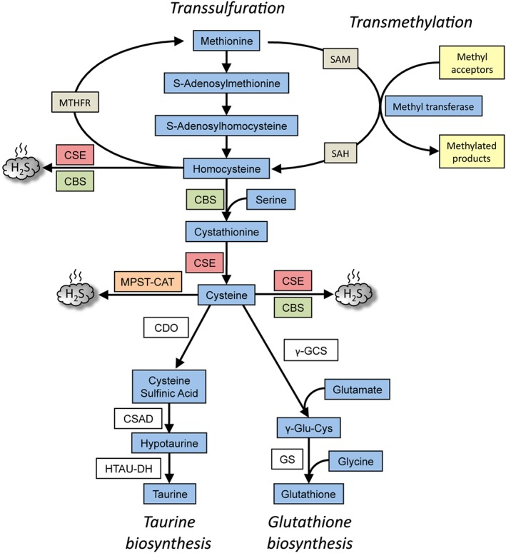
The transsulfuration pathway is a metabolic pathway where transfer of sulfur from homocysteine to cysteine occurs. The pathway leads to the generation of several sulfur metabolites, which include cysteine, GSH and the gaseous signalling molecule hydrogen sulfide (H2 S). Precise control of this pathway is critical for maintenance of optimal cellular function and, therefore, the key enzymes of the pathway, cystathionine β-synthase and cystathionine γ-lyase, are regulated at multiple levels. Disruption of the transsulfuration pathway contributes to the pathology of several conditions such as vascular dysfunction, Huntington's disease and during ageing. Treatment with donors of hydrogen sulfide and/or stimulation of this pathway have proved beneficial in several of these disorders. In this review, we focus on the regulation of the transsulfuration pathway pertaining to cysteine and H2 S, which could be targeted to develop novel therapeutics. LINKED ARTICLES: This article is part of a themed section on Chemical Biology of Reactive Sulfur Species. To view the other articles in this section visit http://onlinelibrary.wiley.com/doi/10.1111/bph.v176.4/issuetoc.


Figures

Figure 1
Figure 1
Overview of the transsulfuration pathway. The pathway results in the generation of cysteine from homocysteine, which in turn, is derived from dietary methionine in mammals. CBS condenses homocysteine with serine to generate cystathionine, the substrate for CSE, to generate cysteine. CSE can generate H2S from either cysteine or homocysteine. While CSE can utilize cysteine to generate H2S, CBS uses a combination of cysteine and homocysteine to generate H2S. A third H2S‐generating enzyme, MPST, in conjunction with cysteine amino acid transferase (CAT) utilizes cysteine to generate H2S. The transsulfuration pathway intersects with the transmethylation pathway at homocysteine, which can be remethylated back to methionine by N5,N10‐methylenetetrahydrofolate reductase (MTHFR). The cysteine generated by the pathway can be chanelled into GSH synthesis by the action of the enzymes, γ‐glutamyl cysteine synthetase (γ‐GCS) and glutathione synthetase (GS) or converted to other sulfur‐containing molecules such as taurine. Taurine is generated by the action of three enzymes, CDO, cysteine sulfinic acid decarboxylase (CSAD) and hypotaurine dehydrogenase (HTAU‐DH).
Figure 2
Figure 2
Regulation of CSE and CBS. CBS and CSE are regulated at multiple levels. (A) Post‐translational regulation of CBS. CBS is a constitutively expressed enzyme and can be regulated by several post‐translational modifications (PTMs) such as sumoylation, phosphorylation and glutathionylation. It is allosterically regulated by SAM, which activates it and increases its stability. The haem centre in CBS can bind NO and CO. (B) CSE is a highly inducible protein, which is regulated by a wide variety of stimuli such as inflammation, ER stress (which can cause translocation to the mitochondria), oxidative stress and Golgi stress, starvation and by hormones. CSE is also modified by PTMs such as phosphorylation, sulfhydration and sumoylation (which may cause translocation to the nucleus). (C) Unlike CBS, whose activity in the cell is regulated predominantly by PTMs, the effects of CSE are exerted mainly by regulation of its expression at the transcriptional level. Several transcription factors, such as the cAMP‐response element binding protein (CREB), SP1, FXR, activating transcription factor 4 and Nrf2, have binding sites on the CSE promoter. In addition, the expression of CSE is also regulated by methylation of its promoter.
Figure 3
Figure 3
Huntington's disease (HD) as an example of disrupted cysteine metabolism. In normal striatal cells, during basal conditions, the expression of CSE is controlled by the transcription factor SP1, which has binding sites in the CSE (Cth) promoter, resulting in cysteine (denoted as C) and GSH production. When cysteine becomes limiting, ATF4 is induced leading to an elevated expression of CSE as well the transporters for cystine (denoted as C–C) (System Xc ). System Xc imports cystine, the oxidized form of cysteine, which is subsequently reduced to cysteine inside cells. Both the basal as well as ATF4‐mediated induction of CSE in response to cysteine deprivation are compromised in HD. SP1 is sequestered by mHtt, leading to diminished expression of CSE, causing an increase in ROS. Initially, ATF4 is functional, but during disease progression, a further increase in ROS prevents this response, leading to a decline in its induction. Stimulating the transsulfuration pathway via the Golgi stress response (as shown in the case of monensin, an ionophore and Golgi stressor) can protect HD cells and prolong survival.
Figure 4
Figure 4
Strategies used to promote redox balance in cells via cysteine metabolism: Huntington's disease as an example. In conditions involving elevated oxidative stress caused by cysteine imbalance (as exemplified by Huntington's disease), several approaches can be followed to improve cell survival. Supplementation of cysteine or N‐acetyl cysteine (NAC) via the diet can reverse abnormalities in cells. Mitigating oxidative stress, which improves stress‐response pathways, can promote optimal functioning of the transsulfuration pathway. Up‐regulating the expression of CSE via the stress‐responsive transcription factor ATF4 can correct abnormalities associated with cysteine deprivation. Mild forms of ER and Golgi stress can elicit cytoprotective responses, which may provoke cellular adaptations that can help protect against future damaging insults. In addition to these strategies, altering the epigenetic state of the CSE and CBS promoters may also induce their expression.

References

    1. Abbott MH, Folstein SE, Abbey H, Pyeritz RE (1987). Psychiatric manifestations of homocystinuria due to cystathionine β‐synthase deficiency: prevalence, natural history, and relationship to neurologic impairment and vitamin B6‐responsiveness. Am J Med Genet 26: 959–969. - PubMed
    1. Agrawal N, Banerjee R (2008). Human polycomb 2 protein is a SUMO E3 ligase and alleviates substrate‐induced inhibition of cystathionine beta‐synthase sumoylation. PLoS One 3: e4032. - PMC - PubMed
    1. Alexander SPH, Cidlowski JA, Kelly E, Marrion NV, Peters JA, Faccenda E et al (2017a). The Concise Guide to PHARMACOLOGY 2017/18: Nuclear hormone receptors. Br J Pharmacol 174: S208–S224. - PMC - PubMed
    1. Alexander SPH, Fabbro D, Kelly E, Marrion NV, Peters JA, Faccenda E et al (2017b). The Concise Guide to PHARMACOLOGY 2017/18: Enzymes. Br J Pharmacol 174: S272–S359. - PMC - PubMed
    1. Alexander SP, Kelly E, Marrion NV, Peters JA, Faccenda E, Harding SD et al (2017c). The Concise Guide to PHARMACOLOGY 2017/18: Overview. Br J Pharmacol 174: S1–S16. - PMC - PubMed

Substances