. 2018 Sep 19;46(16):8616-8629.
doi: 10.1093/nar/gky622.

DNA binding with a minimal scaffold: structure-function analysis of Lig E DNA ligases

Affiliations

DNA binding with a minimal scaffold: structure-function analysis of Lig E DNA ligases

Adele Williamson et al. Nucleic Acids Res. .

Abstract

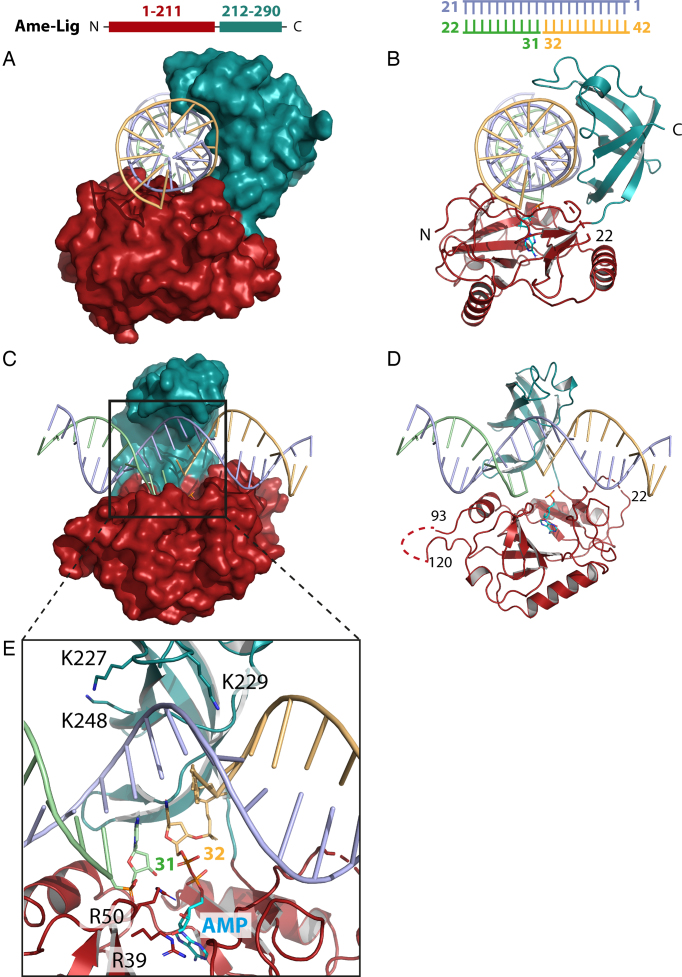
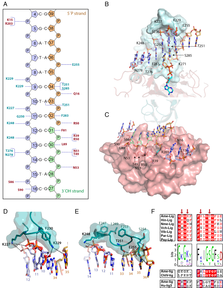
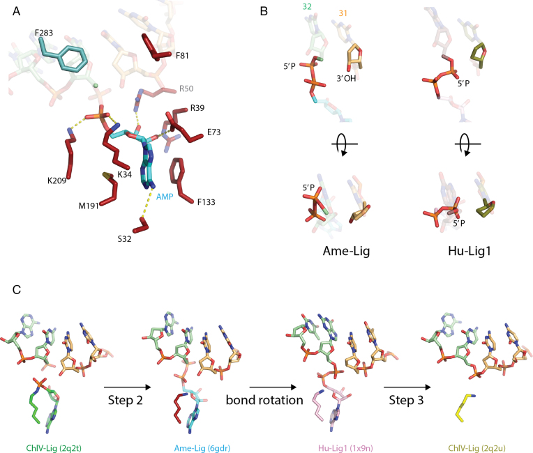
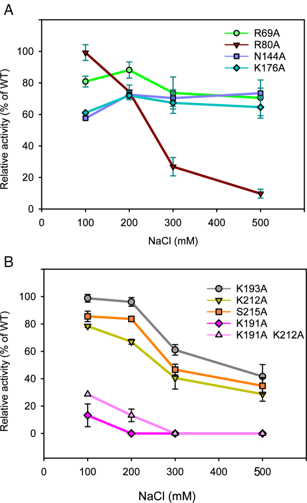
DNA ligases join breaks in the phosphodiester backbone of DNA by catalysing the formation of bonds between opposing 5'P and 3'OH ends in an adenylation-dependent manner. Catalysis is accompanied by reorientation of two core domains to provide access to the active site for cofactor utilization and enable substrate binding and product release. The general paradigm is that DNA ligases engage their DNA substrate through complete encirclement of the duplex, completed by inter-domain kissing contacts via loops or additional domains. The recent structure of a minimal Lig E-type DNA ligase, however, implies it must use a different mechanism, as it lacks any domains or loops appending the catalytic core which could complete encirclement. In the present study, we have used a structure-guided mutagenesis approach to investigate the role of conserved regions in the Lig E proteins with respect to DNA binding. We report the structure of a Lig-E type DNA ligase bound to the nicked DNA-adenylate reaction intermediate, confirming that complete encirclement is unnecessary for substrate engagement. Biochemical and biophysical measurements of point mutants to residues implicated in binding highlight the importance of basic residues in the OB domain, and inter-domain contacts to the linker.


Figures

Figure 1.
Figure 1.
Two views of the overall structure of Ame-Lig bound to 21-bp nicked DNA-adenylate with the protein represented as a surface (A and C) or cartoon showing secondary structural elements (B and D). Detail of the active site is shown in panel E.
Figure 2.
Figure 2.
Ame-Lig binding to DNA. Throughout the figure the text and structure are coloured as follows: the AD-domain red and the OB-domain cyan, the 3′ OH oligo green, the 5′P oligo orange and the complement strand purple. Polar contacts are indicted by dashed grey lines. (A) Schematic of interactions for Ame-Lig predicted by NuProPlot (20). Default settings were applied (maximum distance 3.25 Å, Van der Waals distance 3.80 Å, minimum angle 85°) and confirmed by manual inspection. (B) Interactions of the complement and 5′PO4 strand with the OB domain. (C) Interactions of the nicked and complement strands with residues in the AD domain. (D) DNA interactions of conserved binding motif GKGKF. (E) DNA interactions with conserved motif KLGTG. (F) Conservation of motifs among a selection of aligned Lig Es (right) and sequence logo built from 542 Lig Es (Supplementary Table S2). Corresponding regions of Chlorella virus ligase (ChlV-Lig) and Human Lig 3 (Hu-Lig3) are shown for reference. Naming of sequences is as follows: Hin-lig, Haemophilus influenzae; Nme-lig, Neisseria meningitidis; Vch-lig, Vibrio cholera; Vib-lig, Aliivibrio salomicida; ChlV, Chlorella virus; Hu-lig3, Human ligase3.
Figure 3.
Figure 3.
Active site of Ame-Lig. (A) side-chain interactions with the adenylated nick. AMP is shown in blue, AD-domain side-chain in red, OB-side chains in teal. Hydrogen bonds are indicated by dashed yellow lines. (B) Two views of the phosphodiester bond between the 5′ phosphate of the nick and the AMP highlighting the difference in orientation between the Ame-Lig structure (3′ terminus colored light orange) and Hu-Lig1 (3′ terminus coloured olive). Note that the latter structure was crystalized with a 3′ deoxy nucleotide at the nick terminus to prevent nick joining. (C) Steps of nick-sealing captured in different ligase–DNA complexes by X-ray crystallography. The DNA-bound enzyme adenylate of ChlV-Lig (4), the step 2 intermediates from Ame-Lig and Hu-Lig1 (8) before and efter phosphodiester bond rotation, and ChlV-Lig after step-3 nick sealing before product release.
Figure 4.
Figure 4.
NaCl dependence of Psy-Lig point mutants. Ligase activity is measured by MB assay, each data point is normalized to WT activity at the same NaCl concentration. Measurements are the mean of three replicate experiments; error bars represent the standard deviation from the mean. Raw data, including the NaCl dependence of the WT enzyme are given in Supplementary Figure S3E.
Figure 5.
Figure 5.
Binding affinity of Psy-Lig mutants for nicked DNA. (A) EMSA with FAM-labeled DNA incubated with a 40×, 20× or 10× molar excess of DNA ligase. Bound DNA is shown in the upper panel, free DNA from the same lane in the lower. WT Psy-Lig binding to linear DNA is shown in an adjacent panel. (B) MST binding curves of Psy-Lig AD-domain mutants (left) and OB-domain mutants (right).
Figure 6.
Figure 6.
Comparison of Lig E domains in DNA-bound and apo- conformations. AD-domain is shown in red, OB domain is shown in cyan/blue. Polar contacts are indicted by dashed gray lines. (A) Overall domain conformations. (B) Rearrangement of hydrogen bonding patterns between three conformations. (C) Comparison of hydrophobic interactions between three conformations.
Figure 7.
Figure 7.
Stability of Psy-Lig mutants measured by DSF. In the case that multiple transitions were observed, Tm values are given for the higher temperature, with the exception of K25A which had a single low Tm. (A) Difference in Tm with additives; reference condition is 0.1 mM ATP, 10 mM MgCl2. Asterisks mark conditions where no high-temperature transition was observed. (B) Tm of mutants in buffer C (50 mM Tris pH 8.0, 100 mM NaCl, 1 mM DTT, 5% glycerol) without any additives.

References

    1. Tomkinson A.E., Vijayakumar S., Pascal J.M., Ellenberger T.. DNA ligases: structure, reaction mechanism, and function. Chem. Rev. 2006; 106:687–699. - PubMed
    1. Ellenberger T., Tomkinson A.E.. Eukaryotic DNA ligases: structural and functional insights. Annu. Rev. Biochem. 2008; 77:313–338. - PMC - PubMed
    1. Shuman S. DNA ligases: progress and prospects. J. Biol. Chem. 2009; 284:17365–17369. - PMC - PubMed
    1. Nair P.A., Nandakumar J., Smith P., Odell M., Lima C.D., Shuman S.. Structural basis for nick recognition by a minimal pluripotent DNA ligase. Nat. Struct. Mol. Biol. 2007; 14:770–778. - PubMed
    1. Pitcher R.S., Brissett N.C., Doherty A.J.. Nonhomologous end-joining in bacteria: a microbial perspective. Annu. Rev. Microbiol. 2007; 61:259–282. - PubMed

Publication types

MeSH terms