

Editorial
doi: 10.1007/s12551-018-0444-1.
Epub 2018 Jul 14.
Non-sarcomeric causes of heart failure
Affiliations
- PMID: 30008146
- PMCID: PMC6082313
- DOI: 10.1007/s12551-018-0444-1
Item in Clipboard
Editorial
Non-sarcomeric causes of heart failure
Biophys Rev.
2018 Aug.
No abstract available
Conflict of interest statement
Katja Gehmlich declares that she has no conflicts of interest. Elisabeth Ehler declares that she has no conflicts of interest.
This article does not contain any studies with human participants or animals performed by any of the authors.
Figures
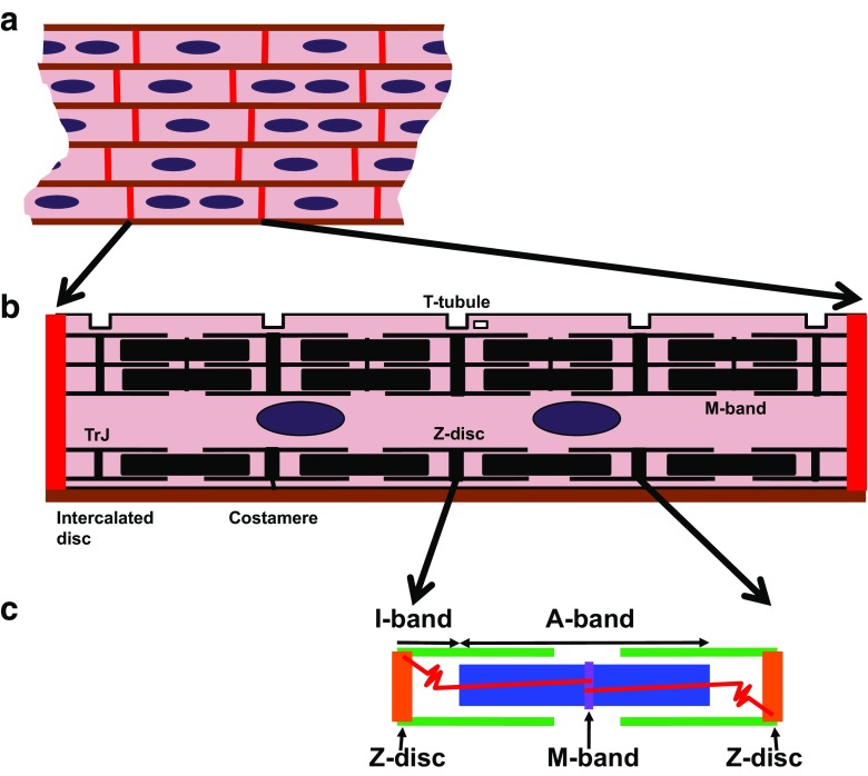
Schematic illustration of heart tissue, a single cardiomyocyte and the sarcomere, the basic unit of a myofibril. a Schematic view of longitudinally sectioned heart tissue (non-muscle cells, such as fibroblasts and endothelial and smooth muscle cells making up blood vessels are not shown). In the murine and human heart the tissue is made up of mono- and binucleated cardiomyocytes; cells are depicted in pink, nuclei in purple, the specialised cell-cell contacts, the intercalated discs are shown in red and the lateral extracellular matrix between the strands of cardiomyocytes is coloured brown. b A simplified scheme of a rod-shaped cardiomyocyte. Myofibrils (not at real scale) are shown in black, intercalated disc in red, nuclei in purple, extracellular matrix in brown. Mitochondria (30% of cell volume) are not depicted. TrJ stands for transitional junction, a kind of “Z-disc light” that links the last sarcomere to the intercalated disc. c A scheme of a sarcomere, the basic unit of a myofibril. It stretches between two Z-discs (shown in orange; marker protein is sarcomeric alpha-actinin), which mark the tranverse borders, in the middle is another transverse structure, the M-band (shown in purple, marker protein is myomesin). The elastic filaments, composed of titin (in red), link the two transverse structures, with titin’s N-terminus being anchored at the Z-discs, while its C-terminus sits in the M-band. The thin filaments (green; main components are actin, tropomyosin, troponin T, I and C) and thick filaments (blue; made up of myosin heavy chain, myosin light chain, myosin binding protein-C) interdigitate and are responsible for sarcomere shortening during contraction
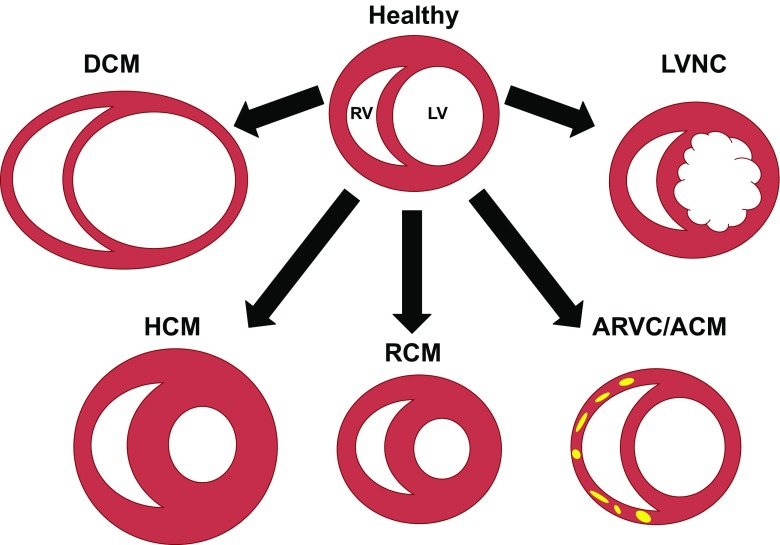
Schematic illustration of different types of cardiomyopathy. A cross-section through the ventricles is shown. Dilated cardiomyopathy (DCM) is characterised by increased ventricular diameter and thinning of the wall, while hypertrophic cardiomyopathy (HCM) shows thickened ventricular and septal walls and reduced left ventricular chamber volume, a feature it shares with restrictive cardiomyopathy (RCM). Arrhythmogenic (right ventricular) cardiomyopathy (ARVC/ACM) can have fibrofatty areas in the RV (shown in yellow), while left ventricular noncompaction cardiomyopathy (LVNC) is characterised by persisting trabeculation and a failure to form compact myocardium. LV left ventricle

Schematic illustration of a single cardiomyocyte. At the bipolar ends are the intercalated discs, the specialised types of cell-cell contacts in the heart. Two myofibrils and two nuclei are shown as well. The approximate subcellular localisation of proteins, cellular structures and mechanistic concepts discussed in the articles that are published in this Special Issue are depicted in red in the cell together with information on the authors. ARVC, arrhythmogenetic right ventricular cardiomyopathy; TrJ, transitional junction; FHL, four and a half LIM domain protein; AS, alternative splicing; RyR, ryanodine receptor; LINC, linker of nucleoskeleton and cytoskeleton; GWAS, genome-wide association studies
References
-
- Agarkova I, Ehler E (2015) The M-band: not just inert glue but playing an active role in the middle of the sarcomere. In: Ehler E (ed) Cardiac cytoarchitecture. How to maintain a working heart, Springer, Cham, pp 125–140
-
- Chong JJ, Yang X, Don CW, Minami E, Liu YW, Weyers JJ, Mahoney WM, Van Biber B, Cook SM, Palpant NJ, Gantz JA, Fugate JA, Muskheli V, Gough GM, Vogel KW, Astley CA, Hotchkiss CE, Baldessari A, Pabon L, Reinecke H, Gill EA, Nelson V, Kiem HP, Laflamme MA, Murry CE. Human embryonic-stem-cell-derived cardiomyocytes regenerate non-human primate hearts. Nature. 2014;510:273–277. doi: 10.1038/nature13233. - DOI - PMC - PubMed
-
- Geier C, Perrot A, Ozcelik C, Binner P, Counsell D, Hoffmann K, Pilz B, Martiniak Y, Gehmlich K, van der Ven PF, Furst DO, Vornwald A, von Hodenberg E, Nurnberg P, Scheffold T, Dietz R, Osterziel KJ. Mutations in the human muscle LIM protein gene in families with hypertrophic cardiomyopathy. Circulation. 2003;107:1390–1395. doi: 10.1161/01.CIR.0000056522.82563.5F. - DOI - PubMed
-
- Hastings R, de Villiers CP, Hooper C, Ormondroyd L, Pagnamenta A, Lise S, Salatino S, Knight SJ, Taylor JC, Thomson KL, Arnold L, Chatziefthimiou SD, Konarev PV, Wilmanns M, Ehler E, Ghisleni A, Gautel M, Blair E, Watkins H, Gehmlich K. Combination of whole genome sequencing, linkage, and functional studies implicates a missense mutation in titin as a cause of autosomal dominant cardiomyopathy with features of left ventricular noncompaction. Circ Cardiovasc Genet. 2016;9:426–435. doi: 10.1161/CIRCGENETICS.116.001431. - DOI - PMC - PubMed
Publication types
Grants and funding
LinkOut - more resources
Full Text Sources
Other Literature Sources
