. 2018 Jul 9;28(13):2103-2114.e5.
doi: 10.1016/j.cub.2018.05.015. Epub 2018 Jun 28.

Marked Diversity of Unique Cortical Enhancers Enables Neuron-Specific Tools by Enhancer-Driven Gene Expression

Affiliations

Marked Diversity of Unique Cortical Enhancers Enables Neuron-Specific Tools by Enhancer-Driven Gene Expression

Stefan Blankvoort et al. Curr Biol. .

Abstract

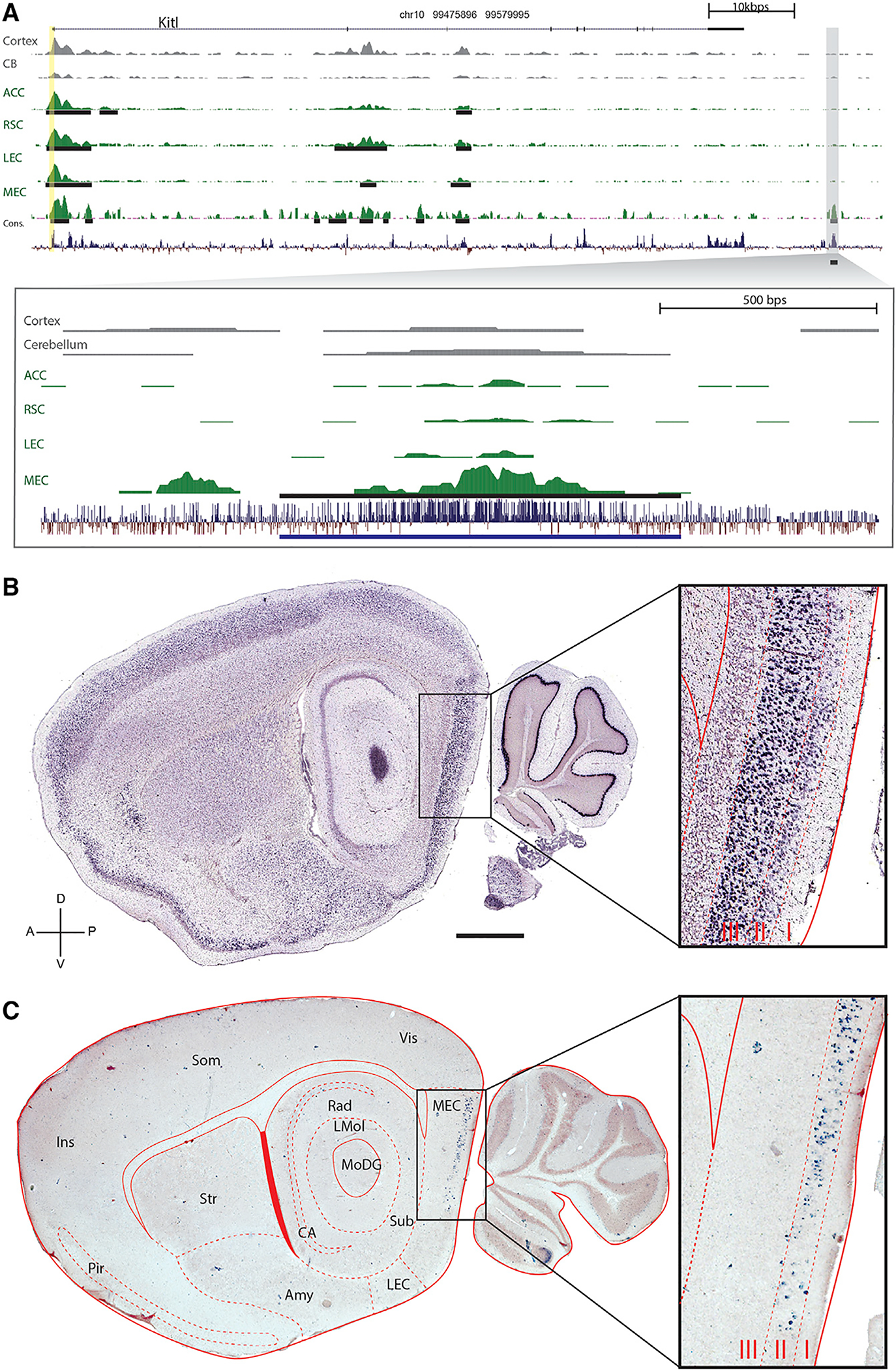
Understanding neural circuit function requires individually addressing their component parts: specific neuronal cell types. However, not only do the precise genetic mechanisms specifying neuronal cell types remain obscure, access to these neuronal cell types by transgenic techniques also remains elusive. Whereas most genes are expressed in the brain, the vast majority are expressed in many different kinds of neurons, suggesting that promoters alone are not sufficiently specific to distinguish cell types. However, there are orders of magnitude more distal genetic cis-regulatory elements controlling transcription (i.e., enhancers), so we screened for enhancer activity in microdissected samples of mouse cortical subregions. This identified thousands of novel putative enhancers, many unique to particular cortical subregions. Pronuclear injection of expression constructs containing such region-specific enhancers resulted in transgenic lines driving expression in distinct sets of cells specifically in the targeted cortical subregions, even though the parent gene's promoter was relatively non-specific. These data showcase the promise of utilizing the genetic mechanisms underlying the specification of diverse neuronal cell types for the development of genetic tools potentially capable of targeting any neuronal circuit of interest, an approach we call enhancer-driven gene expression (EDGE).

Keywords: enhancers; entorhinal cortex; epigenetics; neural circuits; transcriptional control; transgene expression; transgenic animals; transgenic methods; transgenics.


Conflict of interest statement

DECLARATION OF INTERESTS

C.K., J.C., and S.B. are inventors on US Patent Application no. 62/584,282, Appl. Norwegian University of Science and Technology (NTNU), which is related to this work. The authors have no other competing interests to declare.

Figures

Figure 1.
Figure 1.. Experimental Summary of EDGE
(A) Samples of brain regions of interest are microdissected by hand. (B) ChIP-seq is performed on these samples, and genome-wide H3K27ac and H3K4me2 signals for each sample are compared to reference signals and signals from the other samples. Bioinformatic analysis algorithms output unique peaks as potential region-specific enhancers (red bar). (C) Single putative enhancers are cloned into constructs containing a heterologous minimal promoter to drive transgene expression. (D) Following pronuclear injection of these constructs, the resulting founder mice are crossed to reporter lines and evaluated for desired expression patterns. See also Figure S1.
Figure 2.
Figure 2.. ChIP-Seq Reveals a Striking Diversity of Unique and Novel Enhancers in Different Cortical Subregions
(A) Pie charts showing the proportions (and numbers) of distinct active genomic elements identified by H2K27ac ChIP-seq of the 4 cortical subregions. These numbers are roughly similar to those found by ChIP-seq of other organs. (B) Dendrogram (left) and correlation matrix of the H3K27ac signals (right) from replicates of the cortical subregions dissected in this experiment versus those from ENCODE were used for subtraction. Note the relatively high correlation of replicates (except ACC) and clustering of signal from cortical tissues. (C) Heatmaps showing some of the tissue-specific putative enhancers identified in the microdissected cortical subregions. See also Figure S2 and Data S1.
Figure 3.
Figure 3.. The Enhancers of Non-specific Genes Drive Region-Specific Transgene Expression
(A) A genomic view of one of the 165 MEC-specific enhancers yielded by ChIP-seq analysis. The top panel indicates the location and coding regions of Kitl as well as H3K27Ac signal for two regions from roadmap epigenome (cortex and cerebellum), the four regions we analyzed (ACC, RSC, LEC, and MEC), and conservation over 30 species. The vertical yellow column indicates the promoter region upstream of the transcriptional start site. Peak calls are denoted by the black horizontal lines. The specific genomic region containing the enhancer (MEC-13–81) is blown up in the bottom panel. (B) In situ hybridization (ISH) (http://brain-map.org) of Kitl, the gene associated with enhancer MEC-13–81 shows expression throughout cortex, hippocampus, and cerebellum. (C) tTA-dependent transgene Arch driven by the enhancer (ranked number 81) is expressed in MEC LII. The scale bar represents 1,000 μm. Sagittal plane, dorsal-ventral, and anterior-posterior axes are indicated. Amy, amygdala and associated regions; CA, both cornu ammonis fields of the hippocampus; Ins, insular cortex; LEC, lateral entorhinal cortex; LMol, molecular layer of the hippocampus; MEC, medial entorhinal cortex; MoDG, molecular layer of the dentate gyrus; Pir, Piriform cortex; Rad, stratum radiatum of the hippocampus; Som, somatosensory cortex; Str, striatum; sub, subiculum; Vis, visual cortex. Layers I, II, and III of the MEC are indicated in the blow-up to the right. See also Figures S3–S6 and Table S1.
Figure 4.
Figure 4.. Distinct MEC-Specific Enhancers Drive Transgene Expression in Distinct Sets of Cells in MEC
(A–E) ISH showing expression patterns of the native genes Atp10a (A), Odz3 (B), Trps1 (C), Dok5 (D), Nos1 (E) associated with EC-specific enhancers (left column). ISH shows EC-specific expression of transgenes driven by the corresponding EC-specific enhancers (right column); tTA-driven transgenes are in parentheses. ISH for the native genes is from http://brain-map.org. The scale bar in (A) represents 1,000 μm. See also Figures S4–S6 and Table S1.
Figure 5.
Figure 5.. Single Enhancers Can Drive Expression in Histochemically Defined Subsets of MEC LII Cells
(A and B) Horizontal section of a mouse cross between MEC-13–53A and TVAG showing MEC (A) and a full hemisphere (B). Immunohistochemical transgene detection with anti-2A antibody (Ab) shows layer II EC-specific expression. (C, F, and I) Anti-2A histochemistry in sections co-stained against Reelin (C), Calbindin (F), and GAD67 (I). (D) Anti-reelin. (G) Anti-calbindin. (J) Anti-GAD67. (E, H, and K) Overlays of the two signals co-stained against Reelin (E), Calbindin (H), and GAD67 (K); each row is the same section. (L) 100% (1,162/1,162 counted cells) of transgenic cells co-localize with reelin, but there is essentially 0% co-localization with calbindin (2/1,151) and GAD67 (0/738). (M) 49.4% (1,162/2,353) of all reelin-positive cells were positive for the transgene; essentially none of the other cell populations had any transgene-expressing cells. Total numbers of cells are counted in white. (N) Schematic summary of the data in (C)–(M). The scale bars represent 1,000 μm in (B), 200 μm in (A), and 50 μm in (C)–(K). In all graphs, bars show the mean ± SEM. See also Figure S7.
Figure 6.
Figure 6.. Different Single Enhancers Can Drive Expression in Histochemically Distinct Subsets of MEC LII Cells
(A and B) Horizontal section of a mouse cross between MEC-13–104B and tetO-EGFP showing MEC (A) and a full hemisphere (B). Immunohistochemical transgene detection with anti-GFP Ab shows expression in layer II of the EC. (C, F, and I) Anti-GFP histochemistry in sections co-stained against Reelin (C), Calbindin (F), and GAD67 (I). (D) Anti-reelin. (G) Anti-calbindin. (J) Anti-GAD67. (E, H, and K) Overlays of the two signals co-stained against Reelin (E), Calbindin (H), and GAD67 (K); each row is the same section. (L) 43.1% (741/1,717 counted cells) of transgenic cells in layer II of the EC co-localize with reelin whereas 26% (482/1,855) of them co-localize with calbindin. 0% (0/1,579) co-localize with GAD67. (M) 43.1% (741/1,721) of all reelin-positive cells in layer II of the EC were positive for the transgene, and 28.5% (482/1,635) of all calbindin-positive cells in layer II of the EC were positive for the transgene, whereas 0% (0/430) of the GAD67-positive population had any transgene-expressing cells. Total numbers of cells are counted in white. (N) Schematic summary of the data in (C)–(M). The scale bars represent 1,000 μm in (B), 200 μm in (A), and 50 μm in (C)–(K). In all graphs, bars show the mean ± SEM. See also Figure S7.
Figure 7.
Figure 7.. Schematic of Putative Genetic Basis for EDGE Technology
(A) Native gene expression: a gene “X” is expressed in multiple cell types in distinct brain areas. Expression in each cell type is driven by distinct sets of color-coded active enhancers acting upon the native core promoter (pink triangle). Promoter-based methods of transgene expression, such as bacterial artificial chromosome (BAC) transgenesis and knockins, respectively, include several or all of the native enhancers, thereby recapitulating some or all of the expression pattern of the native gene. (B) Enhancer-driven gene expression: a single active enhancer isolated from a particular brain region drives transgene expression from a heterologous minimal promoter (blue). This leads to transgene expression that is restricted to a particular region-specific subset of the cell types that the native promoter expresses in, greatly increasing the anatomical specificity relative to promoter-based methods or the native gene.

References

    1. von Bartheld CS, Bahney J, and Herculano-Houzel S (2016). The search for true numbers of neurons and glial cells in the human brain: A review of 150 years of cell counting. J. Comp. Neurol 524, 3865–3895. - PMC - PubMed
    1. Zeng H, and Sanes JR (2017). Neuronal cell-type classification: challenges, opportunities and the path forward. Nat. Rev. Neurosci 18, 530–546. - PubMed
    1. Bonnavion P, Mickelsen LE, Fujita A, de Lecea L, and Jackson AC (2016). Hubs and spokes of the lateral hypothalamus: cell types, circuits and behaviour. J. Physiol 594, 6443–6462. - PMC - PubMed
    1. Lein ES, Hawrylycz MJ, Ao N, Ayres M, Bensinger A, Bernard A, Boe AF, Boguski MS, Brockway KS, Byrnes EJ, et al. (2007). Genome-wide atlas of gene expression in the adult mouse brain. Nature 445, 168–176. - PubMed
    1. Feng G, Mellor RH, Bernstein M, Keller-Peck C, Nguyen QT, Wallace M, Nerbonne JM, Lichtman JW, and Sanes JR (2000). Imaging neuronal subsets in transgenic mice expressing multiple spectral variants of GFP. Neuron 28, 41–51. - PubMed

Publication types