Dermoscopy as a first step in the diagnosis of onychomycosis
- PMID: 30008642
- PMCID: PMC6041705
- DOI: 10.5114/ada.2018.76220
Dermoscopy as a first step in the diagnosis of onychomycosis
Abstract
Introduction: Over the years, clinical studies have provided new knowledge about the dermoscopic features of the diseases of cutaneous annexes. It seems that dermoscopy has opened a new morphological dimension in the diagnosis and management of hair disorders and onychopathies.
Aim: To identify and describe dermoscopic features of onychomycosis.
Material and methods: A total of 81 consecutive patients with onychomycosis (55 men and 26 women) were prospectively enrolled in the present study. For each patient, all fingernails and toenails were evaluated in clinical and dermoscopic examinations. Mycological tests were performed by potassium hydroxide (KOH) preparation. Mann-Whitney U and χ2 tests were used for the statistical analysis, with a significance threshold of p < 0.05.
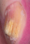
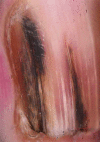
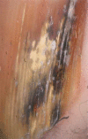
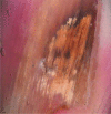
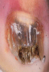
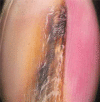
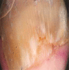
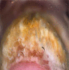
Results: Dermoscopic examination of the patients' nails revealed the following: jagged proximal edge with spikes of the onycholytic area (51.9%), longitudinal streaks and patches (44.4%), subungual hyperkeratosis (27.2%), brown-black pigmentation (9.9%) and leukonychia (1.2%). Jagged proximal edge, subungual hyperkeratosis and leukonychia were positively associated with the onychomycosis type.
Conclusions: Onychomycosis accounts for up to 50% of all consultations for onychopathies. Fast and effective diagnostic approaches are needed in everyday clinical practice. Dermoscopy can provide immediate and accurate information in the diagnosis of onychomycosis. We suggest that dermoscopy should be taken as a first step toward the diagnosis of onychomycosis.
Keywords: dermoscopy; fungal melanonychia; jagged proximal edge; onychomycosis; ruin appearance.
Figures








References
-
- Zalaudek I, Argenziano G, Di Stefani A, et al. Dermoscopy in general dermatology. Dermatology. 2006;212:7–18. - PubMed
-
- Piraccini BM, Bruni F, Starace M. Dermoscopy of non-skin cancer nail disorders. Dermatol Ther. 2012;25:594–602. - PubMed
-
- Scher RK, Tosti A, Joseph WS, et al. Onychomycosis diagnosis and management: perspectives from a Joint Dermatology-Podiatry Roundtable. J Drugs Dermatol. 2015;14:1016–21. - PubMed
LinkOut - more resources
Full Text Sources
Other Literature Sources
