Detection of human papillomavirus in branchial cleft cysts
- PMID: 30008839
- PMCID: PMC6036516
- DOI: 10.3892/ol.2018.8827
Detection of human papillomavirus in branchial cleft cysts
Abstract
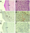
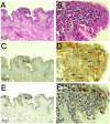
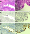
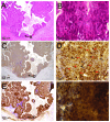
High-risk human papillomavirus (HPV) DNA has been reported to be present in branchial cleft cysts, but further information is required to clarify the role of HPV infection in branchial cleft cysts. The presence of HPV, the viral load and the physical statuses in samples from six patients with branchial cleft cysts were investigated using the polymerase chain reaction (PCR), quantitative PCR, in situ hybridization (ISH) using HPV DNA probes and p16INK4a immunohistochemical analysis. High-risk type HPV-16 DNA was identified in four of the six branchial cleft cysts analyzed. Of the HPV-positive branchial cleft cysts, three exhibited mixed-type integration of HPV. HPV DNA was distributed among the basal-to-granular layers of the cystic wall in ISH analysis, and p16INK4a was weakly expressed in the nuclei and cytoplasm of the same layers in patients with integration. ISH revealed that one patient with episomal-type infection exhibited HPV DNA in the cyst wall and did not express p16INK4a. Two patients without evidence of HPV infection exhibited weak p16INK4a expression in the superficial cyst-lining cells of branchial cleft cysts. These results indicate that infection with high-risk HPV types may be common in branchial cleft cysts. In addition, p16INK4a is not a reliable surrogate marker for HPV infection in branchial cleft cysts.
Keywords: branchial cleft cyst; human papillomavirus; in situ hybridization; p16INK4a expression; viral integration; viral load.
Figures


Similar articles
-
p16(INK4A) immunohistochemical staining may be helpful in distinguishing branchial cleft cysts from cystic squamous cell carcinomas originating in the oropharynx.Cancer. 2009 Apr 25;117(2):108-19. doi: 10.1002/cncy.20001. Cancer. 2009. PMID: 19365840 Free PMC article.
-
Human papillomavirus (HPV) is absent in branchial cleft cysts of the neck distinguishing them from HPV positive cystic metastasis.Acta Otolaryngol. 2018 Sep;138(9):855-858. doi: 10.1080/00016489.2018.1464207. Epub 2018 May 15. Acta Otolaryngol. 2018. PMID: 29764277
-
Lack of evidence of human papillomavirus-induced squamous cell carcinomas of the oral cavity in southern Germany.Oral Oncol. 2013 Sep;49(9):937-942. doi: 10.1016/j.oraloncology.2013.03.451. Epub 2013 Apr 19. Oral Oncol. 2013. PMID: 23608471
-
Combining routine morphology, p16(INK4a) immunohistochemistry, and in situ hybridization for the detection of human papillomavirus infection in penile carcinomas: a tissue microarray study using classifier performance analyses.Urol Oncol. 2014 Feb;32(2):171-7. doi: 10.1016/j.urolonc.2012.04.017. Epub 2013 Mar 14. Urol Oncol. 2014. PMID: 23499169
-
Human papillomavirus infection and immunohistochemical expression of cell cycle proteins pRb, p53, and p16(INK4a) in sinonasal diseases.Infect Agent Cancer. 2015 Aug 4;10:23. doi: 10.1186/s13027-015-0019-8. eCollection 2015. Infect Agent Cancer. 2015. PMID: 26244053 Free PMC article.
Cited by
-
Coordinated Expression of HPV-6 Genes with Predominant E4 and E5 Expression in Laryngeal Papilloma.Microorganisms. 2021 Mar 3;9(3):520. doi: 10.3390/microorganisms9030520. Microorganisms. 2021. PMID: 33802595 Free PMC article.
-
Association between human papillomavirus particle production and the severity of recurrent respiratory papillomatosis.Sci Rep. 2023 Apr 6;13(1):5514. doi: 10.1038/s41598-023-32486-8. Sci Rep. 2023. PMID: 37024540 Free PMC article.
-
Cell-free DNA analysis for recurrent respiratory papillomatosis: A case report.Clin Case Rep. 2024 Aug 6;12(8):e9268. doi: 10.1002/ccr3.9268. eCollection 2024 Aug. Clin Case Rep. 2024. PMID: 39114832 Free PMC article.
-
Human Papillomavirus Infection and EGFR Exon 20 Insertions in Sinonasal Inverted Papilloma and Squamous Cell Carcinoma.J Pers Med. 2023 Apr 11;13(4):657. doi: 10.3390/jpm13040657. J Pers Med. 2023. PMID: 37109043 Free PMC article.
-
Development of Antibodies against HPV-6 and HPV-11 for the Study of Laryngeal Papilloma.Viruses. 2021 Oct 7;13(10):2024. doi: 10.3390/v13102024. Viruses. 2021. PMID: 34696453 Free PMC article.
References
-
- Jacobs MV, Snijders PJ, van den Brule AJ, Helmerhorst TJ, Meijer CJ, Walboomers JM. A general primer GP5+/GP6(+)-mediated PCR-enzyme immunoassay method for rapid detection of 14 high-risk and 6 low-risk human papillomavirus genotypes in cervical scrapings. J Clin Microbiol. 1997;35:791–795. - PMC - PubMed
LinkOut - more resources
Full Text Sources
Other Literature Sources
