. 2018 Jun 12:2018:9405738.
doi: 10.1155/2018/9405738. eCollection 2018.

Moderate Fluid Shear Stress Could Regulate the Cytoskeleton of Nucleus Pulposus and Surrounding Inflammatory Mediators by Activating the FAK-MEK5-ERK5-cFos-AP1 Signaling Pathway

Affiliations

Moderate Fluid Shear Stress Could Regulate the Cytoskeleton of Nucleus Pulposus and Surrounding Inflammatory Mediators by Activating the FAK-MEK5-ERK5-cFos-AP1 Signaling Pathway

Dongping Ye et al. Dis Markers. .

Erratum in

Abstract

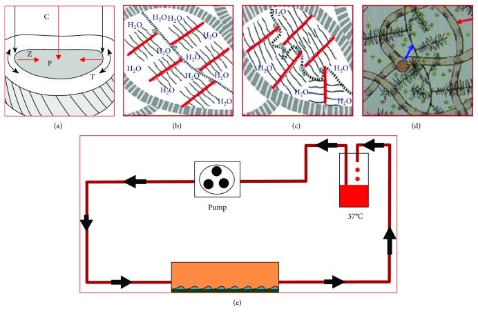
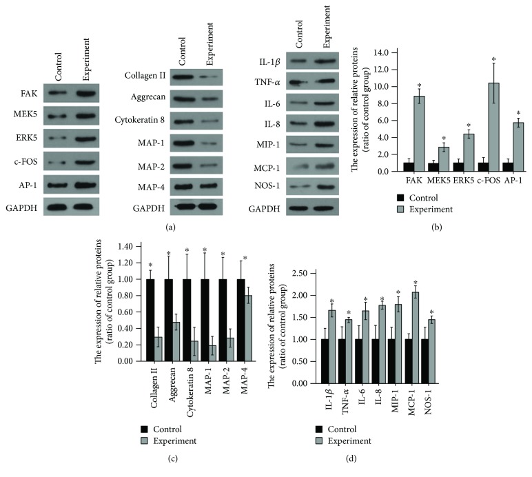
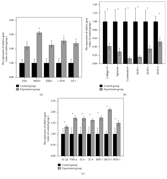
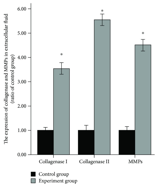
We first applied moderate fluid shear stress to nucleus pulposus cells. The correlation of AP-1 with type II collagen, proteoglycan, Cytokeratin 8 protein, MAP-1, MAP-2, and MAP-4 and the correlation of AP-1 with IL-1β, TNF-α, IL-6, IL-8, MIP-1, MCP-1, and NO were detected. Our results document that moderate fluid shear stress could activate the FAK-MEK5-ERK5-cFos-AP1 signaling pathway. AP1 could downregulate the construct factors of cytoskeleton such as type II collagen, proteoglycan, Cytokeratin 8 protein, MAP-1, MAP-2, and MAP-4 in nucleus pulposus cell after the fluid shear stress was loaded. AP1 could upregulate the inflammatory factors such as IL-1β, TNF-α, IL-6, IL-8, MIP-1, MCP-1, and NO in nucleus pulposus cell after the fluid shear stress was loaded. Taken together, our data suggested that moderate fluid shear stress may play an important role in the cytoskeleton of nucleus pulposus and surrounding inflammatory mediators by activating the FAK-MEK5-ERK5-cFos-AP1 signaling pathway, thereby affecting cell degeneration.


Figures

Figure 1
Figure 1
(a) The structure and function of intervertebral disc: compressive stress (C) produced hydrostatic pressure (P) in nucleus pulposus, then transferred the stress to the fibrous ring, and induced formation of tensile stress (T) in the fibrous ring tissue, induced counterforce of nucleus pulposus by fibrous ring (Z). (b) The proteoglycan was wrapped in a network of collagen fibers (the coarse line of the peripheral zebra). Proteoglycan was composed of a central chain (part of the dotted line) formed by hyaluronic acid. The clustered proteoglycan composed of the core protein (red line) and the glucan sulfate (solid line) was attached to the central chain. (c) Proteoglycan was hydrophilic swelling until pressure balanced with the tensile tension in the collagen fibers, the compress stress on the disc would squeeze part of the water out of the disc, then increased concentration of proteoglycan and increased potential energy of expansion inhibited further compression of nucleus pulposus. Once the compress stress was disappeared, the water returned to the tissue, the new balance was achieved, and the formation of fluid shear stress happened. (d) The blue arrow repressed proteoglycan, and the red arrow repressed collagen fiber. (g) Schematic diagram of fluid shear force device.
Figure 2
Figure 2
(a, b) FAK-MEK5-ERK5-cFos-AP1 pathway-related protein expression obviously increased compared with that before loading. (a, c) The expression of type II collagen, aggrecan, Cytokeratin 8, MAP-1, MAP-2, and MAP-4 proteins noticeably decreased compared with that before loading. (a, d) The expression of inflammatory mediators around nucleus pulposus cells dramatically increased, including IL-1β, TNF-α, IL-6, IL-8, MIP-1, MCP-1, and NO. Repressed p value <0.05, statistical significance was set to α = 0.05 (bilateral). Experiments were repeated three times.
Figure 3
Figure 3
(a) FAK-MEK5-ERK5-cFos-AP1 pathway-related gene expression obviously increased compared with that before loading. (b) The expression of type II collagen, aggrecan, Cytokeratin 8, MAP-1, MAP-2 and MAP-4 mRNA noticeably decreased compared with that before loading. (c) The mRNA expression of inflammatory mediators around nucleus pulposus cells dramatically increased, including IL-1β, TNF-α, IL-6, IL-8, MIP-1, MCP-1 and NO. Repressed p value < 0.05, statistical significance was set to α = 0.05 (bilateral). Experiments were repeated three times.
Figure 4
Figure 4
ELISA results revealed that types I and II collagenase and MMP expression remarkably increased compared with that before loading. Repressed p value < 0.05; statistical significance was set to α = 0.05 (bilateral). Experiments were repeated three times.

Similar articles

Cited by

References

    1. Armbrecht G., Felsenberg D., Ganswindt M., et al. Degenerative inter-vertebral disc disease osteochondrosis intervertebralis in Europe: prevalence, geographic variation and radiological correlates in men and women aged 50 and over. Rheumatology. 2017;56(7):1189–1199. doi: 10.1093/rheumatology/kex040. - DOI - PMC - PubMed
    1. Matta A., Karim M. Z., Isenman D. E., Erwin W. M. Molecular therapy for degenerative disc disease: clues from secretome analysis of the notochordal cell-rich nucleus pulposus. Scientific Reports. 2017;7, article 45623 doi: 10.1038/srep45623. - DOI - PMC - PubMed
    1. Stienen M. N., Smoll N. R., Joswig H., et al. Influence of the mental health status on a new measure of objective functional impairment in lumbar degenerative disc disease. The Spine Journal. 2017;17(6):807–813. doi: 10.1016/j.spinee.2016.12.004. - DOI - PubMed
    1. Canbulat N., Oktenoglu T., Ataker Y., et al. A rehabilitation protocol for patients with lumbar degenerative disc disease treated with posterior transpedicular dynamic stabilization. Turkish Neurosurgery. 2017;27(3):426–435. doi: 10.5137/1019-5149.JTN.16326-15.1. - DOI - PubMed
    1. Grisdela P., Buser Z., D’Oro A., Paholpak P., Liu J. C., Wang J. C. Trends analysis of surgical procedures for cervical degenerative disc disease and myelopathy in patients with tobacco use disorder. European Spine Journal. 2017;26(9):2386–2392. doi: 10.1007/s00586-017-5120-9. - DOI - PubMed

MeSH terms

LinkOut - more resources