

Mandibular Osteitis Leading to the Diagnosis of SAPHO Syndrome
- PMID: 30009075
- PMCID: PMC6020456
- DOI: 10.1155/2018/9142362
Mandibular Osteitis Leading to the Diagnosis of SAPHO Syndrome
Abstract
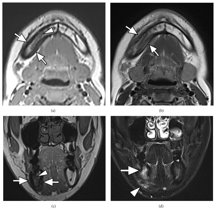
Synovitis, acne, pustulosis, hyperostosis, and osteitis (SAPHO) syndrome is a disorder characterized by pustular skin lesions and osteoarticular lesions. Mandibular involvement of SAPHO syndrome is clinically rare, and it is difficult to reach a diagnosis of SAPHO syndrome from only mandibular manifestations. This report describes the case of a 26-year-old woman who presented with mandibular osteitis. Orthopantomogram and computed tomography showed sclerotic change of the right body of the mandible with periosteal reaction without odontogenic infection, which suggested the possibility of SAPHO syndrome. Detailed medical interview found that she had a history of palmoplantar pustulosis treated at a local dermatology clinic and additional bone scintigraphy showed diffuse increased uptake in the right mandible, as well as in the sternum and the sternocostoclavicular joints. She was eventually diagnosed as having SAPHO syndrome. We should consider SAPHO syndrome when we encounter a patient with mandibular osteitis of unknown etiology.
Figures


References
-
- Chamot A. M., Benhamou C. L., Kahn M. F. The syndrome acne-pustulosis-hyperostosis-osteitis (SAPHO). Results of a national investigation. 85 case-reports. Revue du Rhumatisme et des Maladies Ostéo-Articulaires. 1987;54(3):187–196. - PubMed
-
- Zemann W., Pau M., Feichtinger M., Ferra-Matschy B., Kaercher H. SAPHO syndrome with affection of the mandible: diagnosis, treatment, and review of literature. Oral Surgery, Oral Medicine, Oral Pathology, Oral Radiology, and Endodontology. 2011;111(2):190–195. doi: 10.1016/j.tripleo.2010.04.037. - DOI - PubMed
-
- Karadag-Saygi E., Hakan Gunduz O., Gumrukcu G., Akyuz G. SAPHO syndrome: misdiagnosed and operated. Acta Reumatólogica Portuguesa. 2008;33(4):460–463. - PubMed
-
- Hukuda S., Minami M., Saito T., et al. Spondyloarthropathies in Japan: nationwide questionnaire survey performed by the Japan ankylosing spondylitis society. The Journal of Rheumatology. 2001;28(3):554–559. - PubMed
Publication types
LinkOut - more resources
Full Text Sources
Other Literature Sources
