

The neuropsychological profiles and semantic-critical regions of right semantic dementia
- PMID: 30009130
- PMCID: PMC6041419
- DOI: 10.1016/j.nicl.2018.05.035
The neuropsychological profiles and semantic-critical regions of right semantic dementia
Abstract
Introduction: Previous literature has revealed that the anterior temporal lobe (ATL) is the semantic hub of left-sided or mixed semantic dementia (SD), whilst the semantic hub of right-sided SD has not been examined.
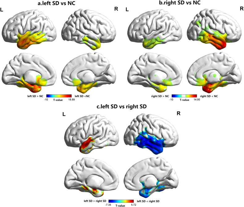
Methods: Seventeen patients with right-sided SD, 18 patients with left-sided SD and 20 normal controls (NC) underwent neuropsychological assessments and magnetic resonance imaging scans. We investigated the relationship between the degree of cerebral atrophy in the whole brain and the severity of semantic deficits in left and right-sided SD samples, respectively.
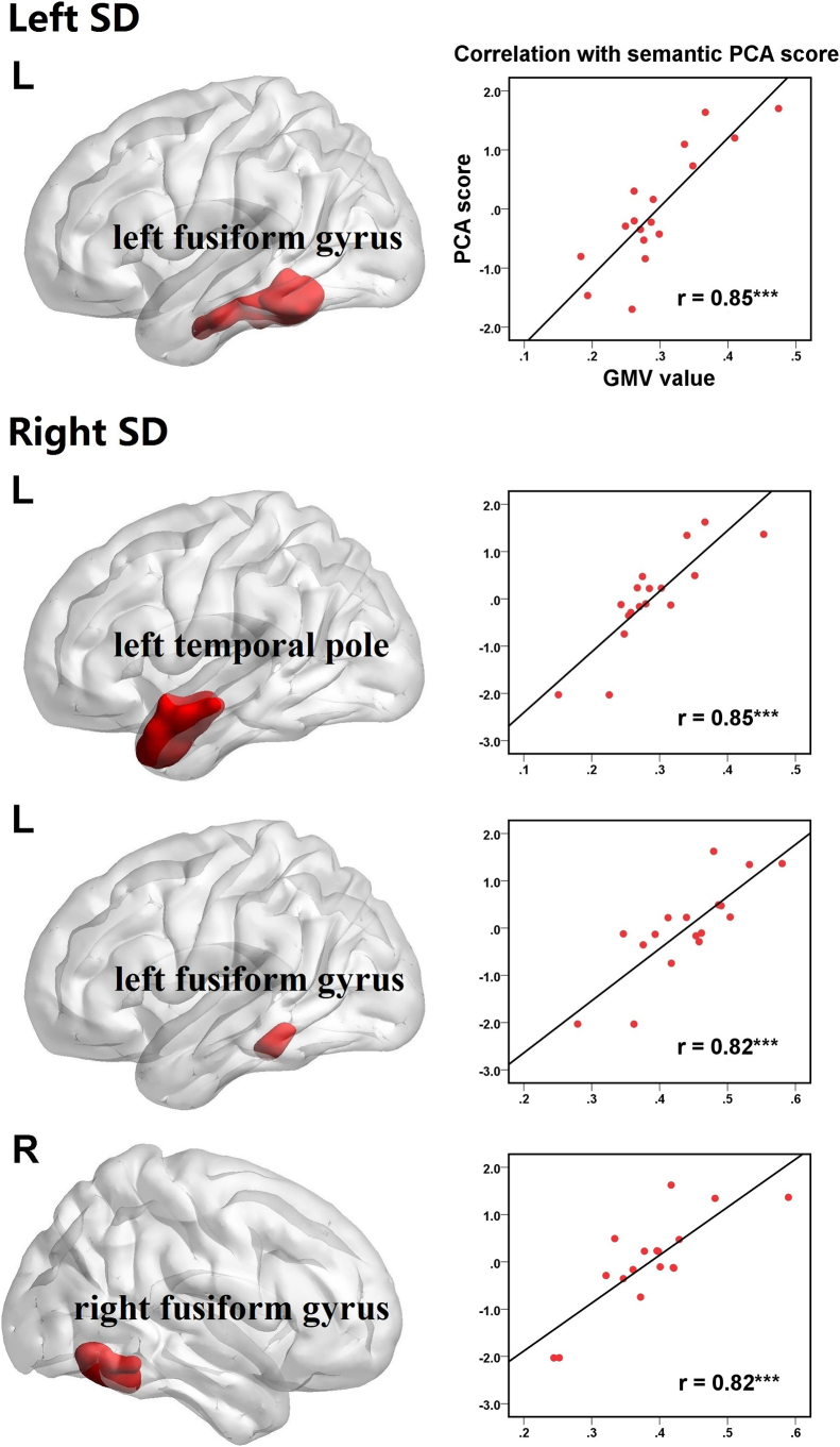
Results: We found the semantic deficits of right-sided SD patients were related to bilateral fusiform gyri and left temporal pole, whilst the left fusiform gyrus correlated with the semantic performance of left-sided SD patients. Moreover, all the findings couldn't be accounted for by total gray matter volume (GMV) or general cognitive degradation of patients.
Discussion: These results provide novel evidence for the current semantic theory, that the important regions for semantic processing include both anterior and posterior temporal lobes.
Keywords: Laterality of brain atrophy; Lesion-behavior mapping; Semantic deficits; Semantic dementia.
Figures
References
-
- Arsalidou M., Taylor M.J. Is 2 + 2 = 4? Meta-analyses of brain areas needed for numbers and calculations. NeuroImage. 2011;54:2382–2393. - PubMed
-
- Bechara A., Damasio H., Damasio A.R. Emotion, decision making and the orbitofrontal cortex. Cereb. Cortex. 2000;10:295–307. - PubMed
-
- Binney R.J., Henry M.L., Babiak M., Pressman P.S., Santos-Santos M.A., Narvid J., Mandelli M.L., Strain P.J., Miller B.L., Rankin K.P., Rosen H.J., Gorno-Tempini M.L. Reading words and other people: a comparison of exception word, familiar face and affect processing in the left and right temporal variants of primary progressive aphasia. Cortex. 2016;82:147–163. - PMC - PubMed
Publication types
MeSH terms
LinkOut - more resources
Full Text Sources
Other Literature Sources
