

Tissue engineering of skin and regenerative medicine for wound care
- PMID: 30009192
- PMCID: PMC6040609
- DOI: 10.1186/s41038-017-0103-y
Tissue engineering of skin and regenerative medicine for wound care
Abstract
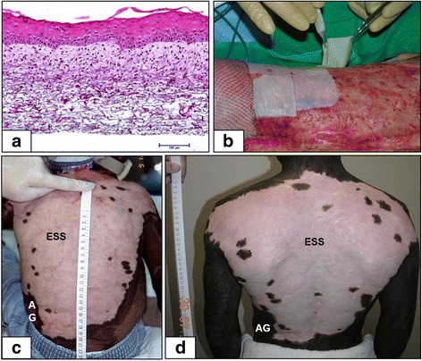
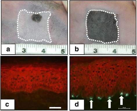
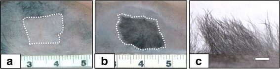
Engineering of biologic skin substitutes has progressed over time from individual applications of skin cells, or biopolymer scaffolds, to combinations of cells and scaffolds for treatment, healing, and closure of acute and chronic skin wounds. Skin substitutes may be categorized into three groups: acellular scaffolds, temporary substitutes containing allogeneic skin cells, and permanent substitutes containing autologous skin cells. Combined use of acellular dermal substitutes with permanent skin substitutes containing autologous cells has been shown to provide definitive wound closure in burns involving greater than 90% of the total body surface area. These advances have contributed to reduced morbidity and mortality from both acute and chronic wounds but, to date, have failed to replace all of the structures and functions of the skin. Among the remaining deficiencies in cellular or biologic skin substitutes are hypopigmentation, absence of stable vascular and lymphatic networks, absence of hair follicles, sebaceous and sweat glands, and incomplete innervation. Correction of these deficiencies depends on regulation of biologic pathways of embryonic and fetal development to restore the full anatomy and physiology of uninjured skin. Elucidation and integration of developmental biology into future models of biologic skin substitutes promises to restore complete anatomy and physiology, and further reduce morbidity from skin wounds and scar. This article offers a review of recent advances in skin cell thrapies and discusses the future prospects in cutaneous regeneration.
Keywords: Burns; Cell therapy; Regenerative medicine; Scar; Skin substitute; Tissue engineering; Wound closure.
Conflict of interest statement
All human subject research was performed with approval of the University of Cincinnati Institutional Review Board with regulatory oversight by the US Food and Drug Administration. All animal subject research was performed with approval of the University of Cincinnati Institutional Animal Care and Use Committee and the Animal Care and Use Research Office of the US Army Medical Research and Materiel Command.An informed consent to publish de-identified data was obtained from the subjects.The authors declare that they have no competing interests.
Figures
References
-
- Finnerty CC, Capek K, Voigt C, Hundeshagen G, Cambiaso-Daniel J, Porter C, Sousse LE, El Ayadi A, Zapata-Sirvent R, Guillory A, et al. The P50 research center in perioperative sciences: how the investment by the National Institute of General Medical Sciences in Team Science has reduced post-burn mortality. J Trauma Acute Care Surg. 2017;83(3):532–542. doi: 10.1097/TA.0000000000001644. - DOI - PMC - PubMed
-
- Boyce ST, Hansbrough JF. Biologic attachment, growth, and differentiation of cultured human epidermal keratinocytes on a graftable collagen and chondroitin-6-sulfate substrate. Surgery. 1988;103:421–431. - PubMed
Publication types
Grants and funding
LinkOut - more resources
Full Text Sources
Other Literature Sources
