

Modified amyloid variants in pathological subgroups of β-amyloidosis
- PMID: 30009199
- PMCID: PMC6043770
- DOI: 10.1002/acn3.577
Modified amyloid variants in pathological subgroups of β-amyloidosis
Abstract
Objective: Amyloid β (Aβ) depositions in plaques and cerebral amyloid angiopathy (CAA) represent common features of Alzheimer's disease (AD). Sequential deposition of post-translationally modified Aβ in plaques characterizes distinct biochemical stages of Aβ maturation. However, the molecular composition of vascular Aβ deposits in CAA and its relation to plaques remain enigmatic.
Methods: Vascular and parenchymal deposits were immunohistochemically analyzed for pyroglutaminated and phosphorylated Aβ in the medial temporal and occipital lobe of 24 controls, 27 pathologically-defined preclinical AD, and 20 symptomatic AD cases.
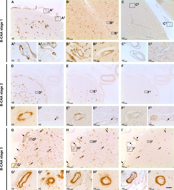
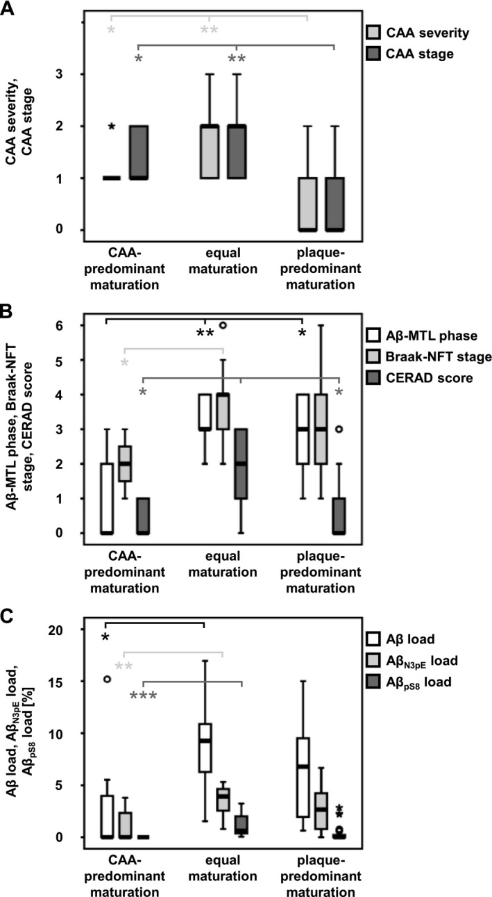
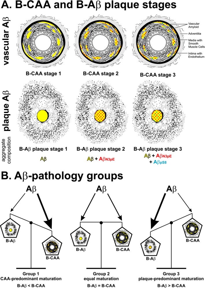
Results: Sequential deposition of Aβ in CAA resembled Aβ maturation in plaques and enabled the distinction of three biochemical stages of CAA. B-CAA stage 1 was characterized by deposition of Aβ in the absence of pyroglutaminated AβN3pE and phosphorylated AβpS8. B-CAA stage 2 showed additional AβN3pE and B-CAA stage 3 additional AβpS8. Based on the Aβ maturation staging in CAA and plaques, three case groups for Aβ pathology could be distinguished: group 1 with advanced Aβ maturation in CAA; group 2 with equal Aβ maturation in CAA and plaques; group 3 with advanced Aβ maturation in plaques. All symptomatic AD cases presented with end-stage plaque maturation, whereas CAA could exhibit immature Aβ deposits. Notably, Aβ pathology group 1 was associated with arterial hypertension, and group 2 with the development of dementia.
Interpretation: Balance of Aβ maturation in CAA and plaques defines distinct pathological subgroups of β-amyloidosis. The association of CAA-related Aβ maturation with cognitive decline, the individual contribution of CAA and plaque pathology to the development of dementia within the defined Aβ pathology subgroups, and the subgroup-related association with arterial hypertension should be considered for differential diagnosis and therapeutic intervention.
Figures
Similar articles
-
Neuropathology and biochemistry of Aβ and its aggregates in Alzheimer's disease.Acta Neuropathol. 2015 Feb;129(2):167-82. doi: 10.1007/s00401-014-1375-y. Epub 2014 Dec 23. Acta Neuropathol. 2015. PMID: 25534025 Review.
-
A distinct brain beta amyloid signature in cerebral amyloid angiopathy compared to Alzheimer's disease.Neurosci Lett. 2019 May 14;701:125-131. doi: 10.1016/j.neulet.2019.02.033. Epub 2019 Feb 23. Neurosci Lett. 2019. PMID: 30807796
-
Amyloid-β plaques may be reduced in advanced stages of cerebral amyloid angiopathy in the elderly.Neuropathology. 2020 Oct;40(5):474-481. doi: 10.1111/neup.12662. Epub 2020 Jun 17. Neuropathology. 2020. PMID: 32557936
-
Origins of Beta Amyloid Differ Between Vascular Amyloid Deposition and Parenchymal Amyloid Plaques in the Spinal Cord of a Mouse Model of Alzheimer's Disease.Mol Neurobiol. 2020 Jan;57(1):278-289. doi: 10.1007/s12035-019-01697-4. Epub 2019 Jul 19. Mol Neurobiol. 2020. PMID: 31325023
-
Cerebral amyloid angiopathy and dementia.Panminerva Med. 2004 Dec;46(4):253-64. Panminerva Med. 2004. PMID: 15876981 Review.
Cited by
-
Amyloid precursor protein-fragments-containing inclusions in cardiomyocytes with basophilic degeneration and its association with cerebral amyloid angiopathy and myocardial fibrosis.Sci Rep. 2018 Nov 9;8(1):16594. doi: 10.1038/s41598-018-34808-7. Sci Rep. 2018. PMID: 30413735 Free PMC article.
-
Insoluble Vascular Amyloid Deposits Trigger Disruption of the Neurovascular Unit in Alzheimer's Disease Brains.Int J Mol Sci. 2021 Apr 1;22(7):3654. doi: 10.3390/ijms22073654. Int J Mol Sci. 2021. PMID: 33915754 Free PMC article.
-
Matrix Development for the Detection of Phosphorylated Amyloid-β Peptides by MALDI-TOF-MS.J Am Soc Mass Spectrom. 2023 Mar 1;34(3):505-512. doi: 10.1021/jasms.2c00270. Epub 2023 Jan 27. J Am Soc Mass Spectrom. 2023. PMID: 36706152 Free PMC article.
-
Phosphorylated Aβ peptides in human Down syndrome brain and different Alzheimer's-like mouse models.Acta Neuropathol Commun. 2020 Jul 29;8(1):118. doi: 10.1186/s40478-020-00959-w. Acta Neuropathol Commun. 2020. PMID: 32727580 Free PMC article.
-
Development of the clinical candidate PBD-C06, a humanized pGlu3-Aβ-specific antibody against Alzheimer's disease with reduced complement activation.Sci Rep. 2020 Feb 24;10(1):3294. doi: 10.1038/s41598-020-60319-5. Sci Rep. 2020. PMID: 32094456 Free PMC article.
References
-
- Selkoe DJ. Alzheimer's disease: genes, proteins, and therapy. Physiol Rev 2001;81:741–766. - PubMed
-
- Attems J, Jellinger K, Thal DR, Van Nostrand W. Review: sporadic cerebral amyloid angiopathy. Neuropathol Appl Neurobiol 2011;37:75–93. - PubMed
-
- Wisniewski H, Johnson AB, Raine CS, et al. Senile plaques and cerebral amyloidosis in aged dogs. A histochemical and ultrastructural study. Lab Invest 1970;23:287–296. - PubMed
-
- Love S, Miners S, Palmer J, et al. Insights into the pathogenesis and pathogenicity of cerebral amyloid angiopathy. Front Biosci (Landmark Ed) 2009;14:4778–4792. - PubMed
LinkOut - more resources
Full Text Sources
Other Literature Sources
