

Altered gene transcription linked to astrocytes and oligodendrocytes in frontal cortex in Creutzfeldt-Jakob disease
- PMID: 30009661
- PMCID: PMC6277179
- DOI: 10.1080/19336896.2018.1500076
Altered gene transcription linked to astrocytes and oligodendrocytes in frontal cortex in Creutzfeldt-Jakob disease
Abstract
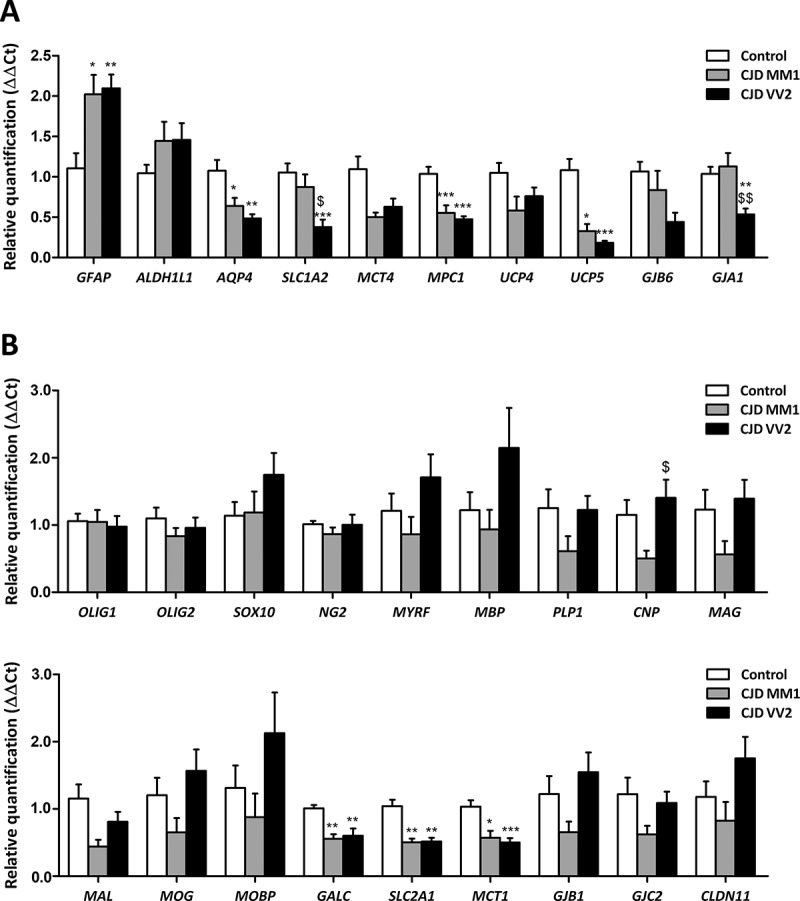
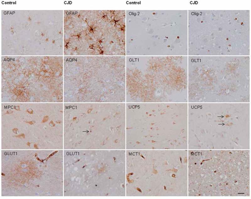
Targeted expression of genes coding for proteins specific to astrocytes, oligodendrocytes and myelin was performed in frontal cortex area 8 of Creutzfeldt-Jakob disease methionine/methionine and valine/valine (CJD MM1 and VV2, respectively) compared with controls. GFAP (glial fibrillary acidic protein) mRNA was up-regulated whereas SLC1A2 (solute carrier family 1 member 2, coding for glutamate transporter 1: GLT1), AQ4 (aquaporin 4), MPC1 (mitochondrial pyruvate carrier 1) and UCP5 (mitochondrial uncoupled protein 5) mRNAs were significantly down-regulated in CJD MM1 and CJD VV2, and GJA1 (connexin 43) in CJD VV2. OLIG1 and OLIG2 (oligodendocyte transcription factor 1 and 2, respectively), SOX10 (SRY-Box10) and oligodendroglial precursor cell (OPC) marker NG2 (neuronal/glial antigen) 2 were preserved, but GALC (coding for galactosylceramidase), SLC2A1 (solute carrier family 2 member 1: glucose transporter member 1: GLUT1) and MCT1 (monocarboxylic acid transporter 1) mRNA expression levels were significantly reduced in CJD MM1 and CJD VV2. Expression levels of most genes linked to myelin were not altered in the cerebral cortex in CJD. Immunohistochemistry to selected proteins disclosed individual variations but GFAP, Olig-2, AQ4 and GLUT1 correlated with mRNA levels, whereas GLT1 was subjected to individual variations. However, MPC1, UCP5 and MCT1 decrease was more closely related to the respective reduced neuronal immunostaining. These observations support the idea that molecular deficits linked to energy metabolism and solute transport in astrocytes and oligodendrocytes, in addition to neurons, are relevant in the pathogenesis of cortical lesions in CJD.
Keywords: Creutzfeldt-Jakob disease; astrocytes; astrogliopathy; energy metabolism; myelin; oligodendrocytes; oligodendrogliopathy; prion diseases.
Figures
References
-
- Prusiner SB. An introduction to prion biology and diseases In: Prusiner SB, editor. Prion biology and diseases, 2nd ed. New York (NY): Cold Spring Harbor Laboratory; 2004. p. 1–87.
-
- Aguzzi A, Sigurdson C, Heikenwaelder M.. Molecular mechanisms of prion pathogenesis. Annu Rev Pathol. 2008;3:11–40. - PubMed
-
- Budka H, Head MW, Ironside JW. Sporadic Creutzfeldt-Jakob disease Dickson DW, Weller RO, et al, editors. Neurodegeneration: the molecular pathology of dementia and movement disorders. 2nd ed. Chichester, West Sussex:Willey-Blackwell; 2011. p.322–335.
-
- Parchi P, Gambetti P, Capellari S. Genetic Creutzfeldt-Jakob disease In: Dickson DW, Weller RO, editors. Neurodegeneration: the molecular pathology of dementia and movement disorders. 2nd ed. Chichester, West Sussex: Willey-Blackwell; 2011. p. 336–345.
Publication types
MeSH terms
Substances
LinkOut - more resources
Full Text Sources
Other Literature Sources
Medical
Miscellaneous