Review
. 2018 Oct 12;430(20):3657-3684.
doi: 10.1016/j.jmb.2018.07.010. Epub 2018 Jul 12.

The Role of Functional Amyloids in Bacterial Virulence

Affiliations
Review

The Role of Functional Amyloids in Bacterial Virulence

Nani Van Gerven et al. J Mol Biol. .

Abstract

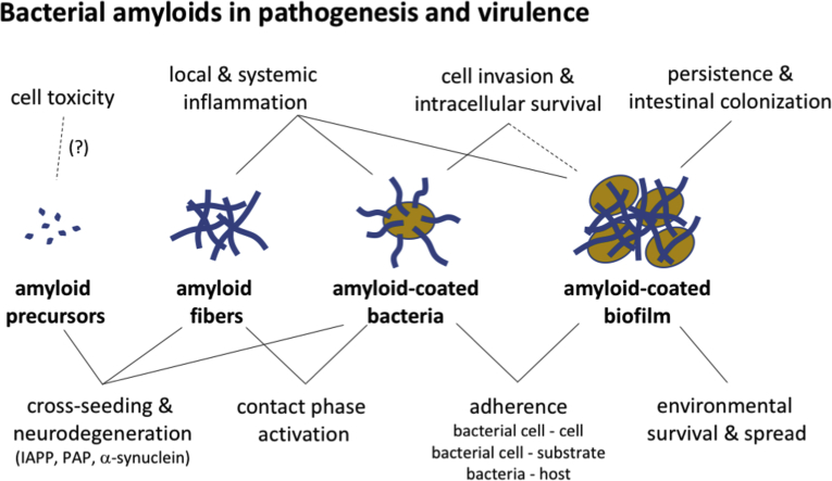
Amyloid fibrils are best known as a product of human and animal protein misfolding disorders, where amyloid formation is associated with cytotoxicity and disease. It is now evident that for some proteins, the amyloid state constitutes the native structure and serves a functional role. These functional amyloids are proving widespread in bacteria and fungi, fulfilling diverse functions as structural components in biofilms or spore coats, as toxins and surface-active fibers, as epigenetic material, peptide reservoirs or adhesins mediating binding to and internalization into host cells. In this review, we will focus on the role of functional amyloids in bacterial pathogenesis. The role of functional amyloids as virulence factor is diverse but mostly indirect. Nevertheless, functional amyloid pathways deserve consideration for the acute and long-term effects of the infectious disease process and may form valid antimicrobial targets.

Keywords: amyloid; bacterial adhesion; bacterial pathogenesis; biofilm; curli.


Figures

Unlabelled Image
Graphical abstract
Fig. 1
Fig. 1
Role of bacterial amyloids in biofilm formation. Representative scanning electron microscopy (SEM) images of curli-expressing E. coli (strain MG1655) show two contributions of curli to biofilm formation: (i) adherence of cells to the underlying substrate, and (ii) formation of an ECM that encapsulates cells and mediates cell–cell contacts in microcolonies and mature biofilms (images reproduced from Ref. [42]). The schematic drawing shows the different stages of biofilm formation, from left to right: initial adhesion to a biotic or abiotic surface, microcolony formation that starts production of a protective ECM, full encapsulation of the colony into a mature 3D biofilm. The contribution of functional amyloids such as curli for adherence and ECM formation is labeled (i) and (ii), respectively. Upper and lower SEM images on the right show the abundant presence of curli or MTP in E. coli or M. tuberculosis biofilms, respectively (images reproduced from Refs. and , respectively). Arrows indicate curli (labeled c) or MTP.
Fig. 2
Fig. 2
Bacterial amyloid assembly pathways. Schematic representation of known bacterial amyloid secretion and assembly pathways, showing the model systems for each of the amyloids. Most systems utilize the generic SEC machinery (SecYEG–SecDF; beige) to transport unfolded units across the cytoplasmic membrane. Some systems encompass pathway-specific transporters (shown in green). In Staphylococci, PSMs are exported via a specialized tetrameric ABC-transporter (PmtA–D), and in Gram-negatives, a dedicated outer membrane channel is required for export to the cell surface, that is, CsgG or FapF in curli and fap, respectively. Several amyloid assembly pathways require accessory proteins (shown in purple), for proteolytic processing (SipW, FapD), export (CsgE, CsgF, FapA) or as a protection against premature amyloid nucleation (CsgC, CsgE). In some systems, minor subunits take a role as nucleator (shown in blue; CsgB, FapB, ChpA–C, TapA), accelerating and localizing the amyloid transition of the major subunit forming the bacterial amyloid fiber (light blue). In Gram-positives, the amyloid fiber can be covalently attached to the peptidoglycan by sortase via the C-terminal LPTX motif. Some proproteins (Bap, P1) are only forming amyloids upon proteolytic processing, and the lighter blue parts indicate protein domains with different folds and functionalities. Where there are clear unfolded intermediates in the amyloid assembly process, these are pictured as random coils in the corresponding colours. Alpha-helical peptides are represented as rods (PSMs, ChpD–H). Genetic organization of the various pathways is drawn with corresponding colors and adjacent genes are indicated were relevant. The curli assembly system is the only system with a known dedicated transcription factor (CsgD; shown in orange). Labels and abbreviations: (DR) Direct Repeat, indicating the bap gene is part of a transposon, encoding its own transposase (Tnp) and putative ABC transporter (ORF1-3). (?) Outstanding questions, (↔) interactions/parallel pathways, (→) unidirectional transport/reactions, (⇄) equilibria, (CslA) cellulose synthase, (IM) cytoplasmic membrane, (OM) outer membrane, (MM) mycomembrane, (PG) peptidoglycan, (AG) arabinogalactan, (C) cellulose. (Inset, upper right) We propose that known bacterial amyloids can be broadly divided into two classes: (I) Intrinsic bacterial amyloids, where the amyloid state is the primary functional form of the proteins involved. These systems appear to have dedicated secretion/assembly pathways. With the exception of chaplins, which form helical precursors, the fiber subunits do not appear to have a folded conformation prior to reaching the amyloid state. (II) Facultative bacterial amyloids are systems where the subunits adopt a (functional) globular folded structure, and can give rise to amyloid fiber formation depending on proteolytic processing and/or environmental triggers such as pH. In these systems, amyloid formation appears to have evolved as a secondary functionality or may be non-functional. In some of these systems, the amyloid state is in equilibrium with free monomers and may act as storage or inactivating state.
Fig. 3
Fig. 3
Transmission electron microscopy images of negatively stained bacteria displaying different bacterial amyloids discussed in this review. From left to right, E. coli cell expressing curli (N. Van Gerven), P. aeruginosa cell expressing fap fibers . M. tuberculosis cell expressing MTP . B. cereus cell expressing TasA fibers , S. aureus cell expressing Bap , or αPSMs in their fibrillar form . Last two images show S. coelicolor expressing chaplins in their adhesive fibrillary form or chaplin/rodlin fibers as part of spore coats (; SEM image).
Fig. 4
Fig. 4
Summarizing scheme of direct and indirect implication of bacterial amyloids in infectious disease and bacterial virulence.

References

    1. Sipe J.D., Cohen A.S. Review: history of the amyloid fibril. J. Struct. Biol. 2000;130:88–98. - PubMed
    1. Eisenberg D.S., Sawaya M.R. Structural studies of amyloid proteins at the molecular level. Annu. Rev. Biochem. 2017;86:69–95. - PubMed
    1. Chiti F., Dobson C.M. Protein misfolding, amyloid formation, and human disease: a summary of progress over the last decade. Annu. Rev. Biochem. 2017;86:27–68. - PubMed
    1. Fowler D.M., Koulov A.V., Balch W.E., Kelly J.W. Functional amyloid—from bacteria to humans. Trends Biochem. Sci. 2007;32:217–224. - PubMed
    1. Larsen P., Nielsen J.L., Dueholm M.S., Wetzel R., Otzen D., Nielsen P.H. Amyloid adhesins are abundant in natural biofilms. Environ. Microbiol. 2007;9:3077–3090. - PubMed

Publication types

MeSH terms