Review
. 2019 Sep:93:36-45.
doi: 10.1016/j.semcdb.2018.07.014. Epub 2018 Sep 5.

Cancer invasion into musculature: Mechanics, molecules and implications

Affiliations
Review

Cancer invasion into musculature: Mechanics, molecules and implications

Lianne Beunk et al. Semin Cell Dev Biol. 2019 Sep.

Abstract

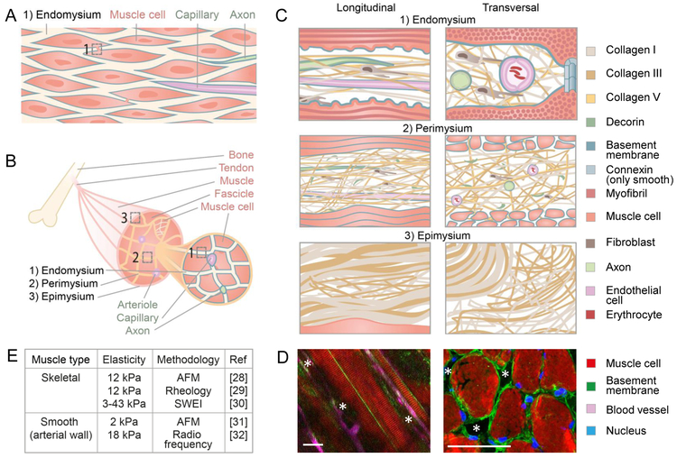
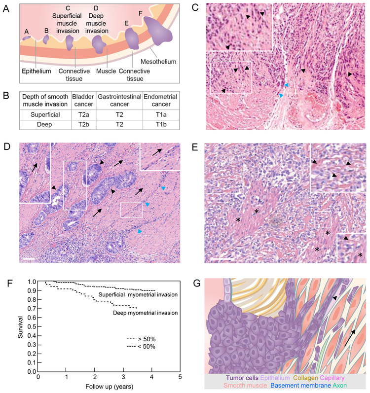
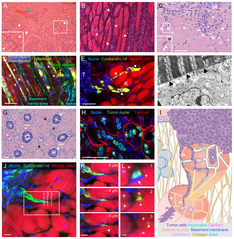
Tumor invasion along structural interphases of surrounding tumor-free tissue represents a key process during tumor progression. Much attention has been devoted to mechanisms of tumor cell migration within extracellular matrix (ECM)-rich connective tissue, however a comprehensive understanding of tumor invasion into tissue of higher structural complexity, such as muscle tissue, is lacking. Muscle invasion in cancer patients is often associated with destructive growth and worsened prognosis. Here, we review biochemical, geometrical and mechanical cues of smooth and skeletal muscle tissues and their relevance for guided invasion of cancer cells. As integrating concept, muscle-organizing ECM-rich surfaces of the epi-, peri- and endomysium provide cleft-like confined spaces along interfaces between dynamic muscle cells, which provide molecular and physical cues that guide migrating cancer cells, forming a possible contribution to cancer progression.

Keywords: Cancer progression; Extracellular matrix; Guided cell migration; Smooth/skeletal muscle tissue; Tumour invasion.


Figures

Figure 1:
Figure 1:. Anatomy of smooth and skeletal muscle tissue.
(A) Smooth muscle tissue consisting of smooth muscle cells connected by gap junctions, covered by a basement membrane (blue) and embedded in collagen-rich endomysium that contains lymph vessels (not shown), blood vessels and axons. (B) Skeletal muscle comprising three structural hierarchical levels: a muscle consisting of muscle bundles (fascicles) which contain muscle cells covered by a basement membrane (blue). (C) The endo-, peri-, and epimysium containing collagen fibrils, fibers, and bundles, respectively, consisting of collagen I, III (and V in the endo- and perimysium only) and basement membranes surrounding muscle cells, axons and blood vessels [16,20,21,23]. All cartoons are magnifications of the numbered regions in (A) and (B). (D) Topology of skeletal muscle in the mouse represented as longitudinal (left) and perpendicular (right) view with asterisks marking anticipated channels. Asterisks, anticipated confined mysial clefts and channels. Scale bars: 50 µm. Left image modified and reprinted with permission from The Company of Biologists; [24]. (E) Elastic moduli of bulk muscle tissue obtained by different methods, including Atomic Force Microscopy (AFM) and Shear Wave Elasticity Imaging (SWEI). Measurements of arterial tissue include besides smooth muscle tissue also other structures, such as elastic fibers in other layers of the arterial wall and endothelia [–32].
Figure 2:
Figure 2:. Overview, histopathology and impact of smooth muscle neoplastic invasion.
(A) Hollow organs such as bladder, gastrointestinal tract and uterus lined by epithelium, connective tissue and smooth muscle layers (muscularis mucosa and muscularis propria) when invaded by tumors (purple) at their onset (A,B), during superficial (C) and deep muscle invasion (D), and when ultimately reaching connective and fatty tissue of nearby organs (E,F). (B) Tumor - Lymph Node - Metastasis (TNM) staging of primary tumors from the indicated neoplasias as defined by the American Joint Committee on Cancer, with T1 defining the smallest, and T4 defining the greatest local extent of the primary tumor. The depth of muscle invasion differentiates tumors into different T-sub-stages, depending on the tumor type [98]. (C) Bladder cancer cell invasion (purple) in chain-like fashion into surrounding smooth muscle tissue (black arrowheads and inset) or along connective tissue (blue arrowheads). Image reprinted with permission from Springer Nature; [40]. (D) Colorectal cancer bud formation (black arrowheads) and immune infiltration (blue arrowheads) along the orientation (arrows; also see insets) of the smooth muscle layer of the colon. (E) Muscle destruction in the anus caused by advanced colorectal cancer disease (also see insets; arrowheads point to remnant muscle structures; asterisks indicate partly remaining muscle). (F) Deep cancer invasion of the myometrium surrounding the uterus is associated with significantly (P <0.0001) decreased overall survival and thus serves as a prognostic parameter. Two hundred and two patients with <50% myometrial invasion and 49 patients with myometrial invasion exceeding 50%. Image modified and reprinted with permission from John Wiley and Sons; [45]. (G) Schematic representation of tumor cell guidance by smooth muscle tissue. Guiding structures for migrating tumor cells (in the direction of the arrow) include endomysium and basement membranes of muscle cells, axons and capillaries as well as the lumen of capillaries (arrowhead). Colored structures are identical to their annotation in Fig. 1A and C. All scale bars, 100 µm.
Figure 3:
Figure 3:. Histopathology of cancer invasion into skeletal muscle tissue.
Examples of skeletal muscle invasion by different human (A,B,C,G) and experimental animal (D,E,F,H) neoplasias. (A) Thyroid cancer cell migration (purple) along the endomysium (white arrowheads) and perimysium (blue arrowheads; also see inset) of skeletal muscle (pink) while muscle integrity remains intact. Image reprinted with permission from Springer Nature; [47]. (B) Non-Hodgkin lymphoma (purple; white arrowheads) infiltrating the endomysium of skeletal muscle tissue (pink; asterisks). Image reprinted with permission from SAGE Publications; [48]. (C) Collective and single muscle invasion of thyroid cancer cells (arrowheads) guided by intact muscle cells (asterisks). See inset for an example of single cell migration between 2 muslce fibers. (D,E,F) High resolution examples of neoplastic invasion along skeletal muscle cells in animal models. (D) Fluorescently labeled murine B16F10 melanoma cells injected into the deep dermis of C57/Bl6 J mice invading into the hypodermis along the pre-existing architecture of the panniculus carnosus muscle (asterisks). Image reprinted with the permission from Landes Bioscience; [12]. (E) Cancer cells, expressing cytokeratin-14 (K) as a basal epithelial marker, migrating as cellular streams (arrowheads) between muscle cells in a transgenic spontaneous breast cancer mouse model. Nuclei on the left side of the image depict cytokeratin-14 negative non-invading tumor cells (T). Image reprinted with permission from Elsevier; [50]. (F) Rat Rd/3 tumor cells injected into rat abdominal wall invading skeletal muscle 6 days post injection. Numerous cytoplasmic processes of a tumor cell (bottom) closely appose to the muscle cell (arrowheads). Imaging by transmission electron microscopy; magnification: 12.600x. Image reprinted with permission from John Wiley and Sons; [42]. (G,H) Intra-muscle fiber invasion by neoplastic cells. (G) Apparent replacement of abdominal muscle cells by malignant glands from colorectal cancer (arrowheads), in the direct vicinity to dystrophic muscle cells (asterisks). (H) Infiltration of laminin-marked panniculus carnosus skeletal muscle fibers by fluorescently labelled human HT1080 fibrosarcoma cells (asterisks) in nude mice. (I) Principles of skeletal muscle invasion. Guidance of tumor cell migration along the endo-, peri- and epimysium and basement membranes sheathing skeletal muscle cells, axons and capillaries (arrowheads), as well as inside muscle cells (asterisk). Colored structures as indicated and identical to their annotation in Fig. 1B and C. (J-L) Guidance of breast cancer invasion by skeletal muscle cells. File-like strand invasion of cytokeratin-14 positive breast cancer cells between skeletal muscle fibers in a transgenic spontaneous breast cancer mouse model (as described in [50]; image set provided by K. Cheung and A. Ewald). (J) Z-projection of whole tumor invasion field into muscle, with rectangle depicting zone for zoom in (K) and three vertical lines for xz projections in (L). (K) A single strand with a highly protrusive leader cell (arrowhead) migrates between individual muscle cells; single cropped z-sections with indicated distances between each other. (L) Orthogonal xz views of breast cancer cell migration (green) being progressively guided by individual muscle cells (asterisks), even on the level of the protrusion in section 3 (white arrowhead). Also note strongly deformed nuclear cross section in section 1 (arrow). Scale bars, 50 µm (C,D,E,G,H); 10 µm (J,K,L); 1 µm (L).

References

    1. Gupta GP, Massagué J, Cancer metastasis: Building a framework, Cell. 127 (2006) 679–695. doi: 10.1016/j.cell.2006.11.001. - DOI - PubMed
    1. Ridley AJ, Schwartz MA, Burridge K, Firtel RA, Ginsberg MH, Borisy G, Parsons JT, Horwitz AR, Cell migration: Integrating signals from front to back, Science (80-. ). 302 (2003) 1704–1709. doi: 10.1126/science.1092053. - DOI - PubMed
    1. Friedl P, Alexander S, Cancer invasion and the microenvironment: Plasticity and reciprocity, Cell. 147 (2011) 992–1009. doi: 10.1016/j.cell.2011.11.016. - DOI - PubMed
    1. van Helvert S, Storm C, Friedl P, Mechanoreciprocity in cell migration, Nat. Cell Biol. 20 (2018) 8–20. doi: 10.1038/s41556-017-0012-0. - DOI - PMC - PubMed
    1. Charras G, Sahai E, Physical influences of the extracellular environment on cell migration, Nat. Rev. Mol. Cell Biol 15 (2014) 813–824. doi: 10.1038/nrm3897. - DOI - PubMed

Publication types

LinkOut - more resources