Review
. 2018 Oct:73:133-148.
doi: 10.1016/j.bbi.2018.07.012. Epub 2018 Jul 17.

Stress and aging act through common mechanisms to elicit neuroinflammatory priming

Affiliations
Review

Stress and aging act through common mechanisms to elicit neuroinflammatory priming

Laura K Fonken et al. Brain Behav Immun. 2018 Oct.

Abstract

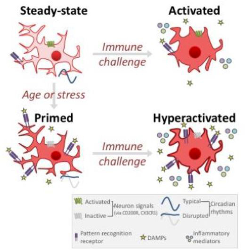
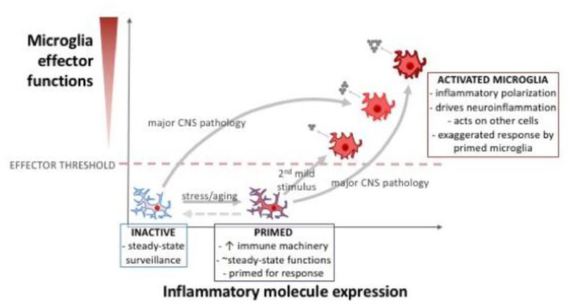
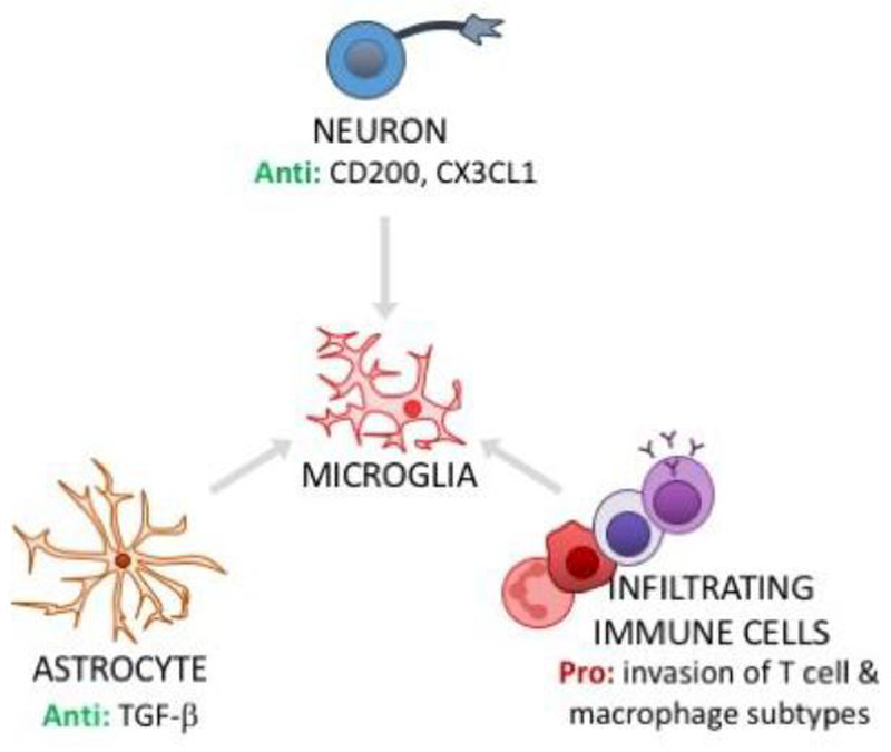
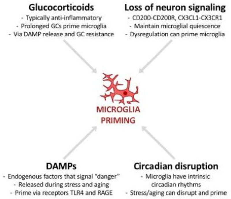
Over the course of an animal's lifespan, there is a protracted breakdown in basic homeostatic functions. Stressors (both psychological and physiological) can accelerate this process and compromise multiple homeostatic mechanisms. For example, both stress and aging can modulate neuroinflammatory function and cause a primed phenotype resulting in a heightened neuroinflammatory profile upon immune activation. Microglia, the brain's resident myeloid cell, produce "silent" immune machinery in response to stress and aging that does not cause immediate immune activation; rather, these changes prime the cell for a subsequent immune insult. Primed microglia exhibit a hyperinflammatory response upon immune activation that can exacerbate pathology. In this review, we will explore parallels between stress- and aging-induced neuroinflammatory priming. First, we will provide a background on the basic principles of neuroimmunology. Next, we will discuss evidence that neuroinflammatory responses become primed in the context of both stress and aging. We will also describe cell-specific contributions to neuroinflammatory priming with a focus on microglia. Finally, common mechanisms underlying priming in the context of stress and aging will be discussed: these mechanisms include glucocorticoid signaling; accumulation of danger signals; dis-inhibition of microglia; and breakdown of circadian rhythms. Overall, there are multifarious parallels between stress- and aging-elicited neuroinflammatory priming, suggesting that stress may promote a form of premature aging. Further unravelling mechanisms underlying priming could lead to improved treatments for buffering against stress- and aging-elicited behavioral pathologies.


Figures

Figure 1.
Figure 1.. Microglial priming during aging and stress.
In the healthy CNS, a steady-state inactive microglial phenotype is associated with limited pattern recognition receptor expression (and low extracellular damage-associated molecular pattern (DAMP) expression), activation of neuron-microglia signaling pathways, and typical daily rhythms in circadian and inflammatory genes. During aging or after stress, microglia develop a “primed” phenotype that does not increase effector function but sensitizes the cells to subsequent stimulation. Primed microglia upregulate pattern recognition receptors, have more DAMPs in the extracellular environment, and de-activated neuron-microglia signaling (dis-inhibition of microglial). When primed microglia experience an immune challenge, the cell responds with an exaggerated inflammatory response that can be prolonged and can exacerbate pathology. Processes shown here represent key common mechanisms driving microglia priming; please see text for more details.
Figure 2.
Figure 2.. Microglial activation occurs along a continuum.
In healthy adult CNS tissue, microglia exist in an inactive state: these microglia survey the CNS microenvironment for infection or damage, and are involved in homeostatic regulation (e.g., synapse modulation, phagocytosis of normal debris, etc.). Following certain physiological challenges (such as stress or aging), microglia can take on a “primed” phenotype. Primed microglia modulate immune machinery involved in an inflammatory response (e.g., increased MHC II, NOD-like receptors, purinergic receptors; see text), but do not substantially increase their inflammatory effector phenotype (i.e., without further stimulus, primed microglia do not release inflammatory molecules). However, if microglia in a primed state are subjected to a stimulus, these primed microglia mount an exaggerated activated response that is maladaptive and can exacerbate pathology. Major CNS pathology (e.g., trauma or ischemia) can directly cause microglia to develop a strongly activated state, but primed microglia may fail to resolve the inflammatory response and continue to produce and secrete inflammatory molecules. By better understanding underlying mechanisms of microglia priming and activation, we may develop effective treatments that dampen aberrant microglial inflammatory phenotypes (dashed arrows).
Figure 3.
Figure 3.. Microglial phenotype is regulated by other cells in the CNS.
In healthy CNS, steady-state microglial quiescence is maintained by signaling with neurons (neuron CD200L to microglia CD200R; neuron CX3CL1 to microglia CX3CR1) and astrocytes (e.g., TGF-β signaling limits microglial activation). Dysregulation of these cell-cell interactions can contribute to microglial priming. In addition, hematogenous immune cells - which typically have no/very limited access to the CNS - can invade CNS tissue in stress and aging. Pro-inflammatory subtypes of infiltrating cells, such as macrophages, can signal to microglia to elicit priming and/or activation. Anti = anti-inflammatory actions; Pro = pro-inflammatory actions.
Figure 4.
Figure 4.. Microglial priming is elicited by common mechanisms in aging and stress.
Microglial priming can be caused by intense and/or prolonged glucocorticoid signaling, by excess release of damage-associated molecular patterns (DAMPs), by loss of neuron signaling (which typically helps maintain microglia homeostasis), and by circadian disruption. Aging and stress can elicit priming by any or all of these mechanisms.

Similar articles

Cited by

References

    1. Abraham J, Johnson RW, 2009. Central inhibition of interleukin-1beta ameliorates sickness behavior in aged mice. Brain Behav Immun 23, 396–401. - PMC - PubMed
    1. Agalave NM, Larsson M, Abdelmoaty S, Su J, Baharpoor A, Lundback P, Palmblad K, Andersson U, Harris H, Svensson CI, 2014. Spinal HMGB1 induces TLR4-mediated long-lasting hypersensitivity and glial activation and regulates pain-like behavior in experimental arthritis. Pain 155, 1802–1813. - PubMed
    1. Ajami B, Bennett JL, Krieger C, Tetzlaff W, Rossi FM, 2007. Local self-renewal can sustain CNS microglia maintenance and function throughout adult life. Nature neuroscience 10, 1538–1543. - PubMed
    1. Alliot F, Godin I, Pessac B, 1999. Microglia derive from progenitors, originating from the yolk sac, and which proliferate in the brain. Brain Res Dev Brain Res 117, 145–152. - PubMed
    1. Askew K, Li K, Olmos-Alonso A, Garcia-Moreno F, Liang Y, Richardson P, Tipton T, Chapman MA, Riecken K, Beccari S, Sierra A, Molnar Z, Cragg MS, Garaschuk O, Perry VH, Gomez-Nicola D, 2017. Coupled Proliferation and Apoptosis Maintain the Rapid Turnover of Microglia in the Adult Brain. Cell Rep 18, 391–405. - PMC - PubMed

Publication types

Substances