. 2018 Aug 1;128(8):3546-3557.
doi: 10.1172/JCI98093. Epub 2018 Jul 16.

Optimal bone fracture repair requires 24R,25-dihydroxyvitamin D3 and its effector molecule FAM57B2

Affiliations

Optimal bone fracture repair requires 24R,25-dihydroxyvitamin D3 and its effector molecule FAM57B2

Corine Martineau et al. J Clin Invest. .

Abstract

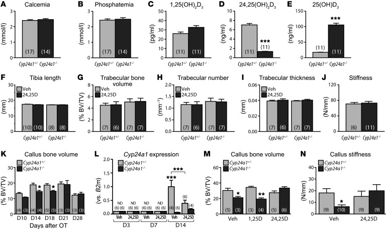
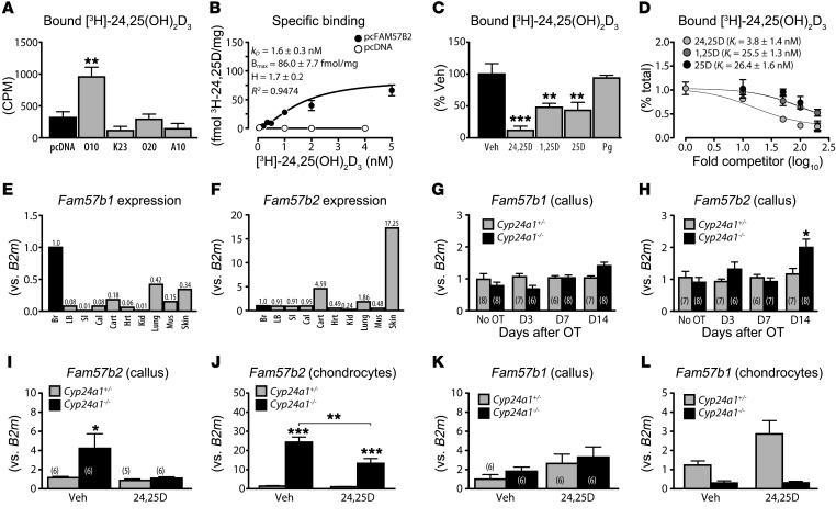
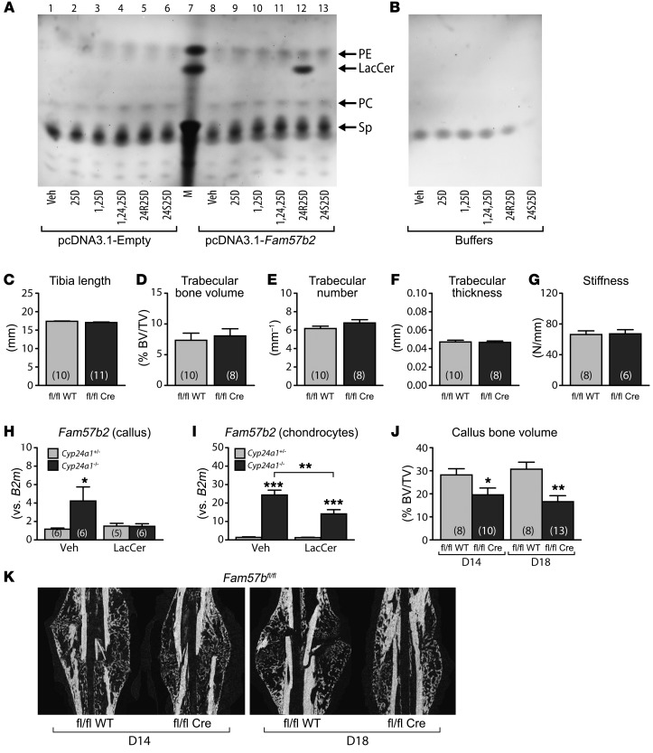
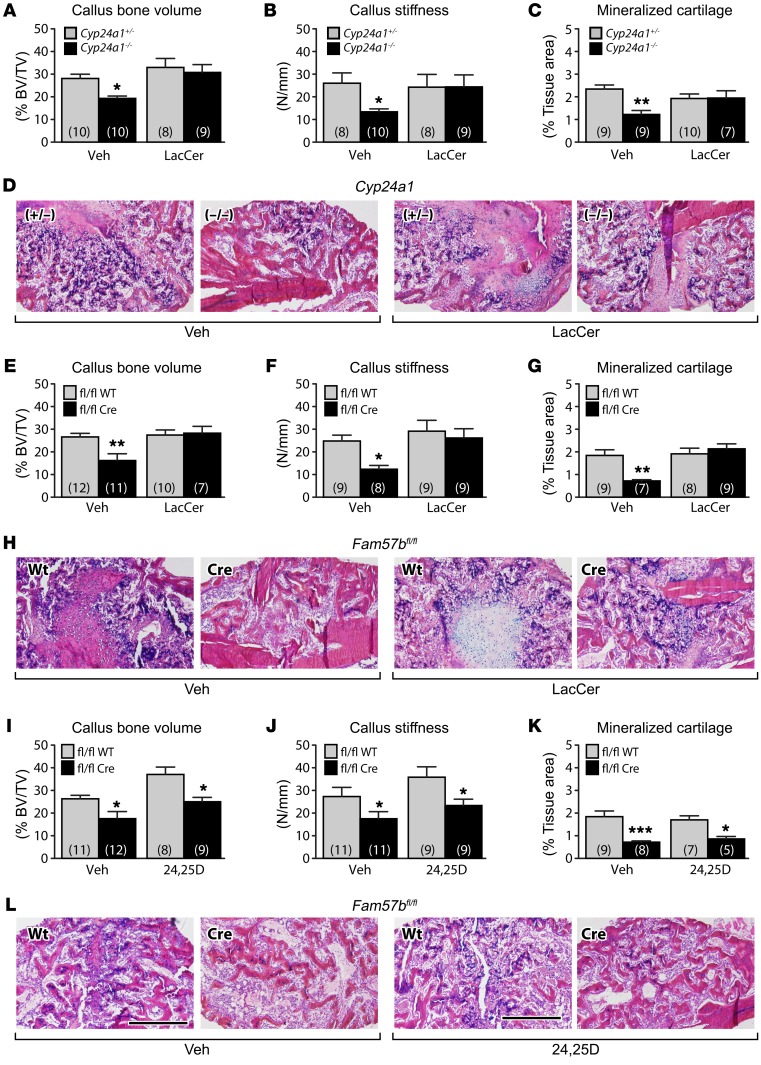
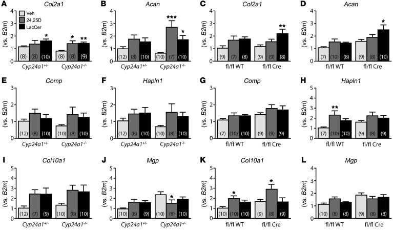
The biological activity of 24R,25-dihydroxyvitamin D3 [24R,25(OH)2D3] remains controversial, but it has been suggested that it contributes to fracture healing. Cyp24a1-/- mice, synthesizing no 24R,25(OH)2D3, show suboptimal endochondral ossification during fracture repair, with smaller callus and reduced stiffness. These defects were corrected by 24R,25(OH)2D3 treatment, but not by 1,25-dihydroxyvitamin D3. Microarrays with Cyp24a1-/- callus mRNA identified FAM57B2 as a mediator of the 24R,25(OH)2D3 effect. FAM57B2 produced lactosylceramide (LacCer) upon specific binding of 24R,25(OH)2D3. Fam57b inactivation in chondrocytes (Col2-Cre Fam57bfl/fl) phenocopied the callus formation defect of Cyp24a1-/- mice. LacCer or 24R,25(OH)2D3 injections restored callus volume, stiffness, and mineralized cartilage area in Cyp24a1-null mice, but only LacCer rescued Col2-Cre Fam57bfl/fl mice. Gene expression in callus tissue suggested that the 24R,25(OH)2D3/FAM57B2 cascade affects cartilage maturation. We describe a previously unrecognized pathway influencing endochondral ossification during bone repair through LacCer production upon binding of 24R,25(OH)2D3 to FAM57B2. Our results identify potential new approaches to ameliorate fracture healing.

Keywords: Bone Biology; Endocrinology; Orthopedics.


Conflict of interest statement

Conflict of interest: The authors have declared that no conflict of interest exists.

Figures

Figure 1
Figure 1. Bone fracture phenotype of Cyp24a1-null mice.
(AE) Blood biochemistry in control and Cyp24a1-mutant mice. (A) Circulating calcium levels. P = 0.4568, by 2-tailed t test. (B) Phosphatemia. P = 0.2429, by 2-tailed t test. (CE) Vitamin D metabolites. ***P < 0.001, by 2-tailed t test. (FI) Static morphometry in untreated and 24R,25(OH)2D3-supplemented control and mutant Cyp24a1 mice. Bone length (F), trabecular bone volume (G), trabecular number (H), and trabecular thickness (I) were assessed by micro-CT. P > 0.05, by 2-way ANOVA followed by Bonferroni’s post test for FI. (J) Bone stiffness was calculated from the 3PBT. P = 0.7420, by 2-tailed t test. (K) Fracture repair callus volume following intramedullary rod–stabilized osteotomy (OT). *P < <0.05, by 2-way ANOVA followed by Bonferroni’s post test. (L) Cyp24a1 expression in callus tissue from control and Cyp24a1-null mice, with or without 24R,25(OH)2D3 rescue. ND, not detectable. ***P < 0.001, by 2-way ANOVA followed by Bonferroni’s post test. (M) Rescue treatment with vitamin D3 metabolites of osteotomies immobilized by external fixator. *P < 0.05 and **P < 0.01, by 2-way ANOVA followed by Bonferroni’s post test. (N) Rescue of biomechanical properties by 24R,25(OH)2D3 administration. *P < 0.05, by 2-way ANOVA followed by Bonferroni’s post test. Data represent the mean ± SEM. The number of animals per group is indicated in parentheses. D, day; 24,25D, 24R,25(OH)2D3; 1,25D, 1,25(OH)2D3; Veh, vehicle.
Figure 2
Figure 2. Cloning and characterization of Fam57b2 from Cyp24a1-null callus tissue.
(A) Binding of [3H]- 24R,25(OH)2D3 to cDNAs identified by microarray analyses. **P < 0.01, by 1-way ANOVA followed by Dunnett’s post test (n = 3). (B) Saturation binding analysis (1-site–specific binding with Hill slope; n = 3). (C and D) Binding competition analysis. **P < 0.01 and ***P < 0.001, by 1-way ANOVA followed by Dunnett’s post test; 1-site fit Ki (n = 3). Pg, progesterone. (E and F) Fam57b isoform expression in tissues. Relative expression levels were determined by reverse transcription quantitative PCR (RT-qPCR) and normalized to B2m. Expression in brain (Br) was arbitrarily assigned a value of 1. LB, long bones; SI, small intestine; Cal, calvaria; Cart, cartilage; Hrt, heart; Kid, kidney; Mus, skeletal muscle. Relative expression is indicated by numerals above each bar. (G and H) Fam57b isoform expression in fracture callus. Relative expression levels were determined at intervals by RT-qPCR and normalized to B2m; expression in contralateral tibia (No OT) was arbitrarily assigned a value of 1. *P < 0.05, by 2-way ANOVA followed by Bonferroni’s post test. (IL) Expression of Fam57b isoforms following 24R,25(OH)2D3 treatment in callus (I and K) or primary cultures of chondrocytes (J and L). *P < 0.05, **P < 0.01, and ***P < 0.001, by 2-way ANOVA followed by Bonferroni’s post test (n = 6). The number of animals per group is indicated in parentheses.
Figure 3
Figure 3. FAM57B2 functional characterization.
(A and B) Ceramide synthase assay. Lysates from HEK293 cells transfected with Fam57b2 or empty vector (A) were used in enzyme assays. Products were separated by TLC as described in Methods. Arrows indicate lipid standards. Results are representative of 3 independent experiments. 25D, 25(OH)D3; 24R25D, 24R,25(OH)2D3; 24S25D, 24S,25(OH)2D3; 1,25D, 1,25(OH)2D3; 1,24,25D, 1,24,25(OH)3D3; M, lipid markers: PE, LacCer, PC, and Sp. (CF) Static morphometry in control (fl/fl WT) and chondrocyte-specific mutant (fl/fl Cre) Fam57b-floxed mice. Bone length (C) and trabecular bone volume (D), number (E), and thickness (F) were assessed by micro-CT. P > 0.05, by 2-tailed t test for CF. (G) Bone stiffness was calculated from the 3PBT. P > 0.05, by 2-tailed t test. (H) Fam57b2 expression in day-18 fracture callus from control or Cyp24a1–/– mice supplemented or not with 50 μg/kg daily C18-LacCer. *P < 0.05, by 2-way ANOVA followed by Bonferroni’s post test. (I) Fam57b2 expression in primary chondrocytes from control or Cyp24a1–/– mice, with or without a 24-hour exposure to 1 μM LacCer. **P < 0.01 and ***P < 0.001, by 2-way ANOVA followed by Bonferroni’s post test (n = 6). (J) Callus volume quantification was determined by micro-CT. *P < 0.05 and **P < 0.01, by 2-way ANOVA followed by Bonferroni’s post test. (K) X-ray projections of callus of chondrocyte-specific Fam57b-deficient mice on day 14 and day 18 after osteotomy. fl/fl WT, control Fam57b mice; fl/fl Cre, chondrocyte-specific Fam57b-deficient mice. The number of animals per group is indicated in parentheses.
Figure 4
Figure 4. Rescue treatment with 24R,25(OH)2D3 or LacCer.
Seventy-two hours after surgery, control (Cyp24a1+/– or fl/fl WT) and mutant (Cyp24a1–/– or fl/fl Cre) mice were subcutaneously injected once daily for fifteen days with vehicle, 6.7 μg/kg 24R,25(OH)2D3 (24,25D), or 50 μg/kg LacCer. Bone volume (A, E, and I) was calculated by micro-CT. Stiffness (B, F, and J) was calculated from 3PBTs. Mineralized cartilage area (C, G, and K) was calculated with ImageJ from histological sections stained with Alcian blue–H&E Y-Orange G trichrome (D, H, and L). *P < 0.05, **P < 0.01, and ***P < 0.001, by 2-way ANOVA followed by Bonferroni’s post test. The number of animals per group is indicated in parentheses. Scale bars: 500 μm (D, H, and L).
Figure 5
Figure 5. Expression monitoring of chondrogenic markers in repair callus.
(AL) RNA was isolated from day-18 repair callus. Relative expression levels of the genes indicated above each panel were determined by RT-qPCR and normalized to B2m. For each gene, expression in vehicle-treated controls was assigned a value of 1. *P < 0.05, **P < 0.01, and ***P < 0.001, by 2-way ANOVA followed by Bonferroni’s post test. The number of animals per group is indicated in parentheses. 24,25D, daily subcutaneous injection of 6.7 μg/kg 24R,25(OH)2D3; LacCer, daily subcutaneous injection with 50 μg/kg C18-LacCer.

Comment in

References

    1. Horst RL, Reinhardt TA, Reddy GS. Vitamin D metabolism. In: Feldman D, Pike JW, Glorieux FH, eds. Vitamin D. 2nd ed. San Diego: Elsevier Academic Press; 2005:15–36.
    1. Sutton AL, MacDonald PN. Vitamin D: more than a “bone-a-fide” hormone. Mol Endocrinol. 2003;17(5):777–791. doi: 10.1210/me.2002-0363. - DOI - PubMed
    1. Makin G, Lohnes D, Byford V, Ray R, Jones G. Target cell metabolism of 1,25-dihydroxyvitamin D3 to calcitroic acid. Evidence for a pathway in kidney and bone involving 24-oxidation. Biochem J. 1989;262(1):173–180. doi: 10.1042/bj2620173. - DOI - PMC - PubMed
    1. Jones G, Prosser DE, Kaufmann M. 25-Hydroxyvitamin D-24-hydroxylase (CYP24A1): its important role in the degradation of vitamin D. Arch Biochem Biophys. 2012;523(1):9–18. doi: 10.1016/j.abb.2011.11.003. - DOI - PubMed
    1. St-Arnaud R. CYP24A1: structure, function, and physiological role. In: Feldman D, Pike JW, Adams JS eds. Vitamin D. 2nd ed. San Diego: Academic Press; 2011:43–56.

Publication types