. 2018 Dec 1;99(6):1171-1183.
doi: 10.1093/biolre/ioy156.

WBP2 shares a common location in mouse spermatozoa with WBP2NL/PAWP and like its descendent is a candidate mouse oocyte-activating factor

Affiliations

WBP2 shares a common location in mouse spermatozoa with WBP2NL/PAWP and like its descendent is a candidate mouse oocyte-activating factor

Lauren E Hamilton et al. Biol Reprod. .

Abstract

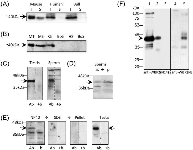
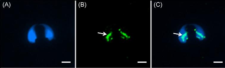
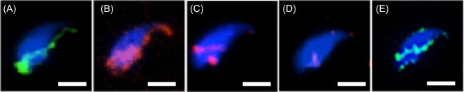
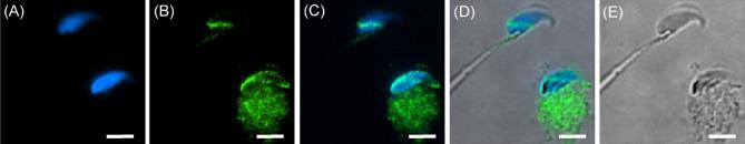
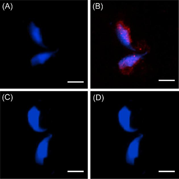
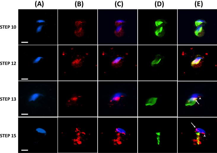
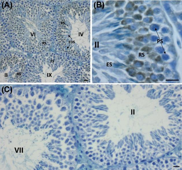
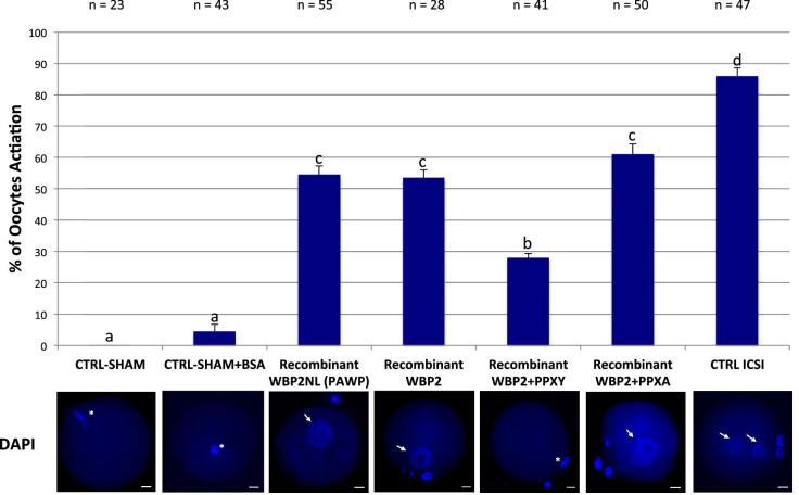
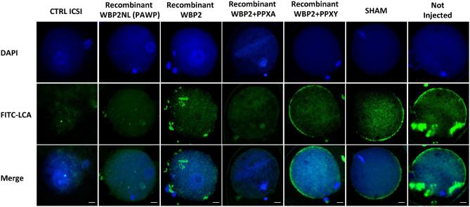
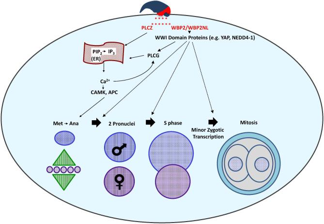
The sperm-borne oocyte-activating factor (SOAF) resides in the sperm perinuclear theca (PT). A consensus has been reached that SOAF most likely resides in the postacrosomal sheath (PAS), which is the first region of the PT to solubilize upon sperm-oocyte fusion. There are two SOAF candidates under consideration: PLCZ1 and WBP2NL. A mouse gene germline ablation of the latter showed that mice remain fertile with no observable phenotype despite the fact that a competitive inhibitor of WBP2NL, derived from its PPXY motif, blocks oocyte activation when coinjected with WBP2NL or spermatozoa. This suggested that the ortholog of WBP2NL, WBP2, containing the same domain and motifs associated with WBP2NL function, might compensate for its deficiency in oocyte activation. Our objectives were to examine whether WBP2 meets the developmental criteria established for SOAF and whether it has oocyte-activating potential. Immunoblotting detected WBP2 in mice testis and sperm and immunofluorescence localized WBP2 to the PAS and perforatorium of the PT. Immunohistochemistry of the testes revealed that WBP2 reactivity was highest in round spermatids and immunofluorescence detected WBP2 in the cytoplasmic lobe of elongating spermatids and colocalized it with the microtubular manchette during PT assembly. Microinjection of the recombinant forms of WBP2 and WBP2NL into metaphase II mouse oocytes resulted in comparable rates of oocyte activation. This study shows that WBP2 shares a similar testicular developmental pattern and location with WBP2NL and a shared ability to activate the oocyte, supporting its consideration as a mouse SOAF component that can compensate for a WBP2NL.


Figures

Figure 1.
Figure 1.
Presence of WBP2 in mouse testis and sperm. (A) WBP2 labeling of a 10% SDS polyacrylamide gel transferred to PVDF. Anti-WBP2 (N-14) antibody labels a band around 40kDa (arrow) in the testis (T) of mouse, human and bull. This band is also present in the mouse sperm (S), but not human or bull spermatozoa. (B) Comparison of the presence of WBP2 in the spermatozoa of different mammalian species. Anti-WBP2 (ProteinTech) antibody labels a band of ∼40 kDa (arrow) in mouse testis (MT), and mouse (MS) and rat spermatozoa (RS); however, the band is not present in boar (BoS), human (HS) or bull (BuS) spermatozoa. Each lane, except the MT, was loaded with 7.5 million spermatozoa. All dashed lines in this and subsequent figures represent cuts from adjacent lanes of the same western membranes. The adjacent lanes in subsequent figures were loaded with the same sample but after transfer, they were incubated under different conditions, usually with the addition of a blocking peptide. (C) The immunoreactivity of the 40kDa band (arrow) in both mouse testis and spermatozoa is absent when the anti-WBP2 antibody (Ab) is preincubated with the WBP2 (N-14) blocking peptide (+b). (D) Comparison of relative content of WBP2 (arrow) in sonicated mouse sperm supernatant (ss) and resulting sperm pellet (p) after separation by centrifugation. (E) western blot showing that WBP2 (arrow) is completely extractable from mouse spermatozoa when incubated in nonionic detergent (NP-40). Subsequent extraction in SDS of the NP-40 extracted sperm pellet was devoid of WBP2 as well as its sperm pellet. Testis was used as a positive control and marker. Ab, Anti-WBP2 (N-14) antibody; +b, antibody plus specific blocking peptide. (F) Comparison of anti-WBP2 (N14) and anti-WBP2NL antibody labeling on westerns of mouse sperm NP-40 extracts and NP-40 extracted sperm pellets. Lane 1; Control mouse testis labeled with anti-WBP2 antibody. Lanes 2 and 4: Mouse sperm NP-40 extracts incubated with anti-WBP2 and anti-WBP2NL antibodies, respectively. Lanes 3 and 5: NP-40 extracted sperm pellets incubated with anti-WBP2 and anti-WBP2NL antibodies, respectively. Note there is no immunocrossreactivity detected between these antibodies. Migration levels of molecular mass standards are denoted on the side in kDa.
Figure 2.
Figure 2.
Mouse spermatozoa extracted from vas deferens that were sonicated prior to fixation in 2% formaldehyde, without permeabilization. (A) DAPI alone. (B) Antibody alone. (C) Merge A and B. Note that under these preparatory conditions the PAS (white arrow) is immunoreactive to the antibody but the perforatorium is not labeled. Blue = DAPI, Green = anti-WBP2 (N-14), bar = 5 μm.
Figure 3.
Figure 3.
Immunocytochemistry showing specificity of WBP2 labeling in mature mouse spermatozoa. Mouse spermatozoa from the vas deferens were sonicated for 5 s, fixed in 4% paraformaldehyde and permeabilized with Triton-X-100. (A) DAPI alone. (B) Antibody alone. (C) Merge A and B. Note in B and C that with the added permeabilization step the perforatorium became immunoreactive (white arrows) in addition to the PAS (compare with Figure 2). (D) When the anti-WBP2 antibody was pre-incubated with its blocking peptide before primary antibody incubation no labeling was found in the PT regions. Blue = DAPI, Red = anti-WBP2 (N-14), bar = 5 μm.
Figure 4.
Figure 4.
Mouse sonicated sperm showing the labeling pattern of WBP2NL (A) and WBP2 (B) when sonicated for 5 s before fixation and permeabilization. When sonicated for a longer time (3 × 10 s) the amount of WBP2 present is greatly reduced (C) but the labeling pattern remains consistent. If the sonicated sperm heads (SSpH) are treated with 2% Triton-X-100 for 2 h at room temperature before fixation and permeabilization the WBP2 labeling is lost (D) as contrary to WBP2NL, which is retained (E). Blue = DAPI, Red or green = anti-WBP2 (N-14), bar = 5 μm.
Figure 5.
Figure 5.
Immunocytochemistry showing the presence of WPB2 in elongating and elongated mouse spermatids fixed with 2% formaldehyde. (A) DAPI alone. (B) Antibody alone. (C) Merge A and B. (D) Merge C with E, which is a differential interference contrast picture of the same field. Note that in the elongated spermatid, at the top of the panel, WBP2 has been incorporated as part of the PAS while in the elongating spermatid, found below, WBP2 is still distributed throughout the cytoplasm lobe. Blue = DAPI, Green = anti-WBP2 (N-14), bar = 5 μm.
Figure 6.
Figure 6.
Confirmation that the WBP2 immunoreactivity in the elongating/elongated spermatid cytoplasm is specific. (A) DAPI alone. (B) DAPI plus anti-WBP2 (N-14) antibody labeling. (C) DAPI alone (D) DAPI and anti-WBP2 (N-14) antibody plus its blocking peptide (compare B to D). Blue = DAPI, Red = anti-WBP2 (N-14), bar = 5 μm.
Figure 7.
Figure 7.
Immunofluorescence showing developmental progression, during the 16 steps of spermiogenesis, in and the association of WBP2 and Tubulin (manchette) in mouse spermatids. Mouse spermatids from testicular extract were fixed in 4% paraformaldehyde and permeabilized after fixation with Triton-X-100. (A) DAPI alone. (B) Anti-WBP2 antibody alone. (C) Merge A and B. (D) Anti-Tubulin antibody alone. (E) C and D merged. Blue = DAPI, Red = anti-WBP2 (N-14), Green = anti-Tubulin, Yellow = the colocalization of WBP2 and Tubulin, Bar = 5 μm.
Figure 8.
Figure 8.
Immunoperoxidase labeling of the mouse seminiferous epithelium with anti-WBP2 (N-14) antibody. Stages of the 12 stage cycle of the mouse seminiferous epithelium are marked by Roman numerals. (A) Low magnification section through mouse seminiferous tubules labeled with anti-WBP2 antibody. Note that the round spermatids (RS) are intensely immunoreactive. (B) Higher magnification of seminiferous epithelium in stage II showing relatively intense immunoreactivity in the cytoplasm of round spermatids compared to elongating spermatids (ES) in step 14 of spermiogenesis and pachytene spematocytes (PS). (C) When the anti-WBP2 antibody was pre-incubated with its blocking peptide before primary antibody incubation no labeling was found anywhere throughout the seminiferous epithelium. Bar in A = 20 μm, Bars in B and C = 10 μm.
Figure 9.
Figure 9.
Comparison of mouse oocyte activation rates between sham, sham + BSA, recombinant (r)WBP2NL (PAWP), rWBP2, rWBP2 + PPXY, rWBP2 + PPXA and sperm heads (CTRL ICSI) 9–10 h after microinjection into metaphase II arrested oocytes (three or more replicates were done for each experimental group except for sham (without BSA) where only 2 replicates were done). In the positive control (ICSI), metaphase II arrested oocytes that were injected with sperm heads had an average activation rate of 86%, based on the formation of male and female pronuclei. Recombinant WBP2NL (0.0075 μg/μl) and rWBP2 (0.0075 μg/μl) injections produced average activation rates over 50%, based on the formation of a female pronucleus. Coinjection of PPXY peptide with rWBP2 significantly reduced the activation rate obtained with rWBP2 alone, while coinjection PPXA peptide with rWBP2 had no significant effect. The negative control, sham + BSA, had a 4% activation rate with most oocytes remaining arrested in metaphase II while sham had a 0% activation rate. Representative DAPI stained images of pronuclei progression within oocytes or ova for respective microinjection regime are found directly below bar graphs. Pronuclei, arrows; metaphase II plate, asterisk; bar = 20 μm. Superscript letters (a, b, c) indicate significant differences at P < 0.05 and error bars denote standard error.
Figure 10.
Figure 10.
Cortical granule exocytosis in rWBP2NL and rWBP2 microinjected mouse oocytes that display pronuclear formation. The pronuclear development in each treatment group was evaluated through nuclear staining (DAPI). The cortical granule exocytosis reaction was assessed through staining with FITC conjugated LCA (FITC-LCA) and both DAPI and FITC-LCA staining were overlaid to show the correlating trends in staining patterns when oocytes were and were not activated (Merge). Bar = 20 μm
Figure 11.
Figure 11.
Proposed two-factor mechanism of sperm-induced oocyte activation. The sperm oocyte-activating factor (SOAF) is released from the PAS region of the sperm head PT into the ooplasm upon sperm–oocyte fusion. The two candidate SOAF proteins, PLCZ1 and WBP2NL/WBP2 would facilitate the hydrolysis of PIP2 to IP3. Next, IP3 would bind to the IP3R receptor on the endoplasmic reticulum (ER), and trigger the release of Ca2+ from its stores, ultimately increasing the intracellular Ca2+ in the oocyte cytosol. This mechanism would be activated either directly, in the case of PLCZ1, or indirectly in the case of WBP2NL/WBP2. It is proposed that WBP2NL/WBP2 would bind to the ooplasmic WWI domain containing proteins to activate oocyte PLC Gamma (PLCG), catalyzing the hydrolysis of PIP2 to IP3. The increase in Ca2+ would result in the downstream activation of enzymes such as the calcium-calmodulin kinase (CAMK) and the anaphase promoting complex (APC) catalyzing the completion of oocyte meiosis and further, repetitive Ca2+-induced Ca2+ release (Ca2+ oscillations). Additionally, WBP2NL/WBP2 may aid in the regulation and/or activation of WWI domain containing proteins shown to have roles in the early stages of zygotic development after the initial activation of the oocyte. Specifically, WBP2/WBP2NL could serve as adaptors of yet to be identified transcription factors responsible for the minor zygotic genome activation, resulting in transcription of a small number of genes in the pronuclei, observed in mammalian zygotes.

Similar articles

Cited by

References

    1. Stice SL, Robl JM. Activation of mammalian oocytes by a factor obtained from rabbit sperm. Mol Reprod Dev 1990; 25:272–280. - PubMed
    1. Swann K. A cytosolic sperm factor stimulates repetitive calcium increases and mimics fertilization in hamster eggs. Development 1990; 110:1295–1302. - PubMed
    1. Swann K, Lai FA. Egg activation at fertilization by a soluble sperm protein. Physiol Rev 2016; 96:127–149. - PubMed
    1. Hachem A, Godwin J, Ruas M, Lee HC, Ferrer BM, Ardestani G, Bassett A, Fox S, Navarrete F, de SP, Heindryckx B, Fissore R et al. . PLCzeta is the physiological trigger of the Ca2+ oscillations that induce embryogenesis in mammals but conception can occur in its absence. Development 2017; 144:2914–2924. - PMC - PubMed
    1. Kimura Y, Yanagimachi R, Kuretake S, Bortkiewicz H, Perry AC, Yanagimachi H. Analysis of mouse oocyte activation suggests the involvement of sperm perinuclear material. Biol Reprod 1998; 58:1407–1415. - PubMed