Review
. 2018 Oct 1;39(5):803-829.
doi: 10.1210/er.2018-00020.

Circulating Testosterone as the Hormonal Basis of Sex Differences in Athletic Performance

Affiliations
Review

Circulating Testosterone as the Hormonal Basis of Sex Differences in Athletic Performance

David J Handelsman et al. Endocr Rev. .

Abstract

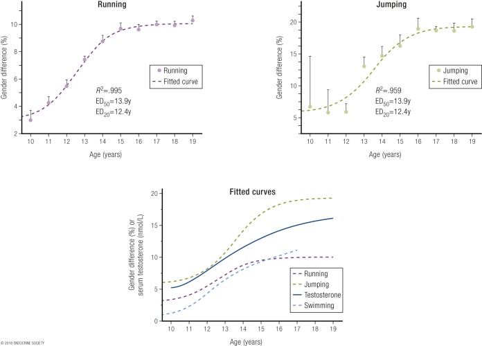
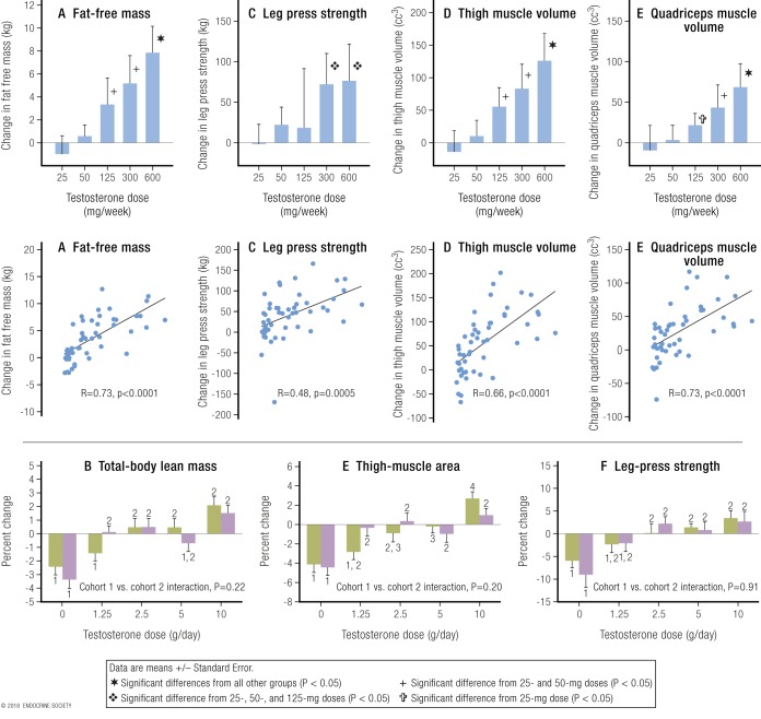
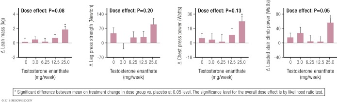
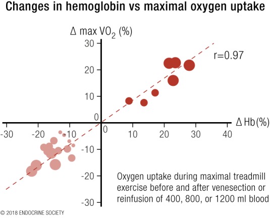
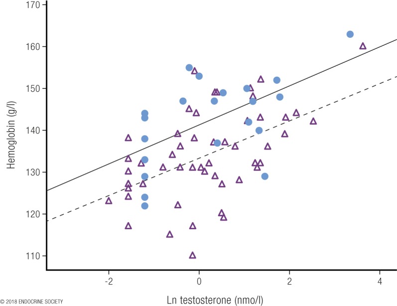
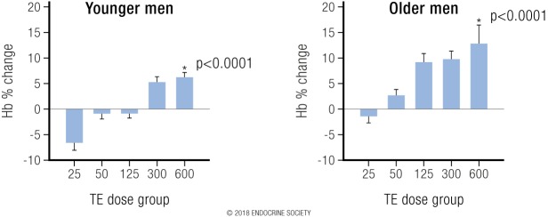
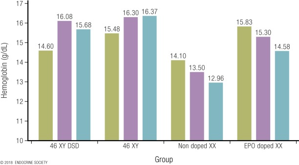
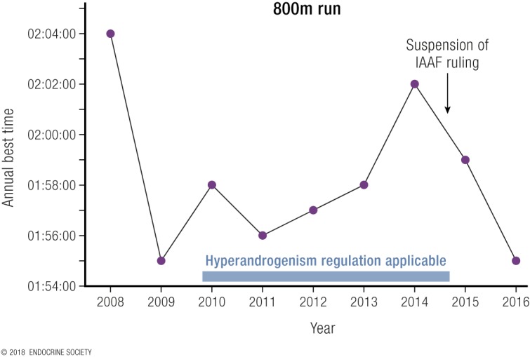
Elite athletic competitions have separate male and female events due to men's physical advantages in strength, speed, and endurance so that a protected female category with objective entry criteria is required. Prior to puberty, there is no sex difference in circulating testosterone concentrations or athletic performance, but from puberty onward a clear sex difference in athletic performance emerges as circulating testosterone concentrations rise in men because testes produce 30 times more testosterone than before puberty with circulating testosterone exceeding 15-fold that of women at any age. There is a wide sex difference in circulating testosterone concentrations and a reproducible dose-response relationship between circulating testosterone and muscle mass and strength as well as circulating hemoglobin in both men and women. These dichotomies largely account for the sex differences in muscle mass and strength and circulating hemoglobin levels that result in at least an 8% to 12% ergogenic advantage in men. Suppression of elevated circulating testosterone of hyperandrogenic athletes results in negative effects on performance, which are reversed when suppression ceases. Based on the nonoverlapping, bimodal distribution of circulating testosterone concentration (measured by liquid chromatography-mass spectrometry)-and making an allowance for women with mild hyperandrogenism, notably women with polycystic ovary syndrome (who are overrepresented in elite athletics)-the appropriate eligibility criterion for female athletic events should be a circulating testosterone of <5.0 nmol/L. This would include all women other than those with untreated hyperandrogenic disorders of sexual development and noncompliant male-to-female transgender as well as testosterone-treated female-to-male transgender or androgen dopers.


Figures

Figure 1.
Figure 1.
Sex differences in performance (in percentage) according to age (in years) in running events, including 50 m to 2 miles (upper left panel), and in jumping events, including high jump, pole vault, triple jump, long jump, and standing long jump (upper right panel) [for details, see Ref. (8)]. The lower panel is a fitted sigmoidal curve plot of sex differences in performance (in percentage) according to age (in years) in running, jumping, and swimming events, as well as the rising serum testosterone concentrations from a large dataset of serum testosterone of males. Note that in the same dataset, female serum testosterone concentrations did not change over those ages, remaining the same as in prepubertal boys and girls. Data are shown as mean and SEM of the pooled sex differences by age. Reproduced with permission from Handelsman DJ. Sex differences in athletic performance emerge coinciding with the onset of male puberty. Clin Endocrinol (Oxf). 2017;87:68–72.
Figure 2.
Figure 2.
Strong dose-response relationship between testosterone dose and circulating concentration with muscle mass and strength in men. The upper panels [from Bhasin et al. (111)] display the strong dose-response relationships of muscle mass shown as (A) “lean” or “fat-free” mass or volume of (D) thigh and (E) quadriceps muscle and (C) of leg muscle strength with increasing testosterone dose (upper row) or circulating concentration (middle row). Serum testosterone concentrations are in US units (ng/dL; divide by 28.8 to get nmol/L). Adapted with permission from Bhasin S, Woodhouse L, Casaburi R, et al. Testosterone dose-response relationships in healthy young men. Am J Physiol Endocrinol Metab. 2001;281:E1172–E1181. The lower panels [from Finkelstein et al. (65)] show the strong dose-response relationships of (B) whole-body muscle mass, (E) thigh muscle mass, and (F) leg press strength with increasing testosterone dose. Cohorts 1 and 2 were treated with the same increasing doses of testosterone but either without (green fill, cohort 1) or with (purple fill, cohort 2) an aromatase inhibitor (anastrozole), which prevents conversion of testosterone to estradiol. The differences between cohorts (i.e., use of anastrozole) was not significant for muscle mass and strength and can be ignored with results of the two cohorts being pooled. Reproduced with permission from Finkelstein JS, Lee H, Burnett-Bowie SA, Pallais JC, et al. Gonadal steroids and body composition, strength, and sexual function in men. N Engl J Med 2013;369:1011–1022.
Figure 3.
Figure 3.
From Huang et al. (112): Dose-response effects on lean (muscle) mass and three measures of muscle strength as a result of increasing doses of weekly testosterone enanthate injections in women. Note the effects on all four parameters (three statistically significant) of the highest testosterone dose, the only one that produced circulating testosterone levels exceeding the normal female range. Reproduced with permission from Huang G, Basaria S, Travison TG, et al. Testosterone dose-response relationships in hysterectomized women with or without oophorectomy: effects on sexual function, body composition, muscle performance and physical function in a randomized trial. Menopause 2014;21:612–623.
Figure 4.
Figure 4.
Redrawn results from Ekblom et al. (124). Results from the transfusion of additional blood are shown in dark red circles and those after blood withdrawal in light red circles. Adapted with permission from Ekblom B, Goldbarg AN, Gullbring B. Response to exercise after blood loss and reinfusion. J Appl Physiol 1972;33:175–180.
Figure 5.
Figure 5.
Plot of circulating hemoglobin against the natural logarithm of serum testosterone in women with congenital adrenal hyperplasia [from Karunasena et al. (92)]. The filled circles represent a cohort where serum testosterone was measured by immunoassay. The open triangles denote a second cohort, where serum testosterone was measured by LC-MS. Note the systematic overestimation of testosterone by the immunoassay used in cohort 1 vs LC-MS measurement in cohort 2. Despite that overestimation, however, the correlations were similar in both cohorts. Reproduced under a Creative Commons BY-NC-ND 4.0 license from Karunasena N, Han TS, Mallappa A, et al. Androgens correlate with increased erythropoiesis in women with congenital adrenal hyperplasia. Clin Endocrinol (Oxf) 2017;86:19–25.
Figure 6.
Figure 6.
From Coviello et al. (131): Depicts the strong dose-response relationship between increasing testosterone dose with resulting change in blood hemoglobin in young and older men. Reproduced with permission from Coviello AD, Kaplan B, Lakshman KM, et al. Effects of graded doses of testosterone on erythropoiesis in healthy young and older men. J Clin Endocrinol Metab 2008;93:914–919.
Figure 7.
Figure 7.
Mean hemoglobin concentrations (g/dL) of 12 elite athletes in 4 groups of 3 XY or XX middle-distance runners. The hemoglobin concentrations were collected as a part of the Athlete Biological Passport and analyzed according to the World Anti-Doping Agency standard methods. Each bar (athlete) is the mean of a minimum of three blood samples. In the 46,XY DSD group, blood was collected in a period when the athlete was not undergoing hormonal suppressive treatment.
Figure 8.
Figure 8.
Best annual 800-m times of an elite female athlete between 2008 and 2016. Data provided by Dr. Richard Auchus, University of Michigan, Ann Arbor, Michigan.

References

    1. Handelsman DJ. Performance enhancing hormones in sports doping In: DeGroot LJ, Jameson JL, eds. Endocrinology. 7th ed.Philadelphia, PA: Elsevier Saunders; 2015:441–454.
    1. Coleman DL. Sex in sport. Available at: ssrn.com/abstract=2928106. Accessed 22 October 2017.
    1. Lee PA, Nordenström A, Houk CP, Ahmed SF, Auchus R, Baratz A, Baratz Dalke K, Liao LM, Lin-Su K, Looijenga LH III, Mazur T, Meyer-Bahlburg HF, Mouriquand P, Quigley CA, Sandberg DE, Vilain E, Witchel S; Global DSD Update Consortium . Global disorders of sex development update since 2006: perceptions, approach and care [published correction appears in Horm Res Paediatr. 2016;85(3):180]. Horm Res Paediatr. 2016;85(3):158–180. - PubMed
    1. Southren AL, Tochimoto S, Carmody NC, Isurugi K. Plasma production rates of testosterone in normal adult men and women and in patients with the syndrome of feminizing testes. J Clin Endocrinol Metab. 1965;25(11):1441–1450. - PubMed
    1. Horton R, Tait JF. Androstenedione production and interconversion rates measured in peripheral blood and studies on the possible site of its conversion to testosterone. J Clin Invest. 1966;45(3):301–313. - PMC - PubMed