. 2019 Jan 15;35(2):319-328.
doi: 10.1093/bioinformatics/bty620.

AuTom-dualx: a toolkit for fully automatic fiducial marker-based alignment of dual-axis tilt series with simultaneous reconstruction

Affiliations

AuTom-dualx: a toolkit for fully automatic fiducial marker-based alignment of dual-axis tilt series with simultaneous reconstruction

Renmin Han et al. Bioinformatics. .

Abstract

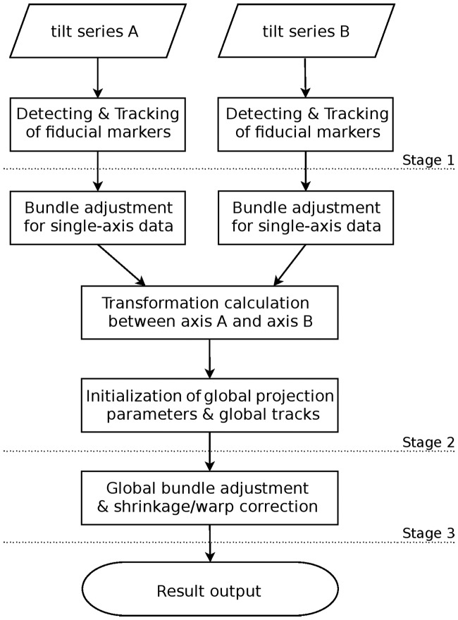
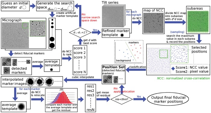
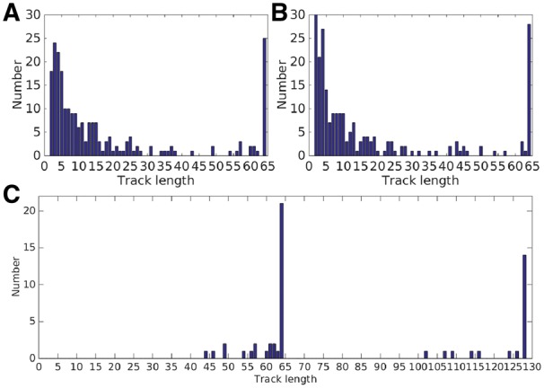
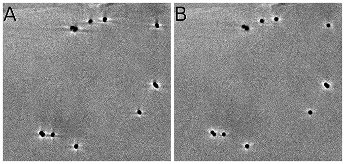
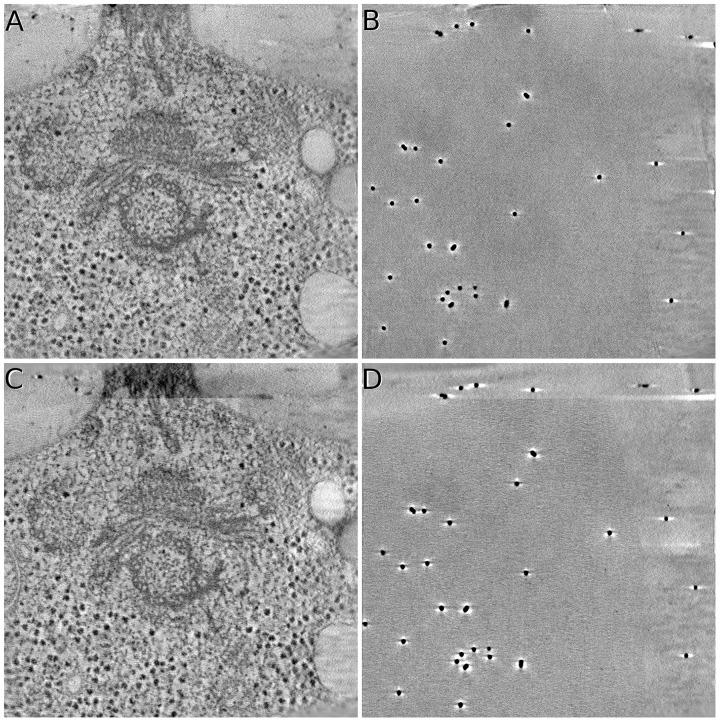
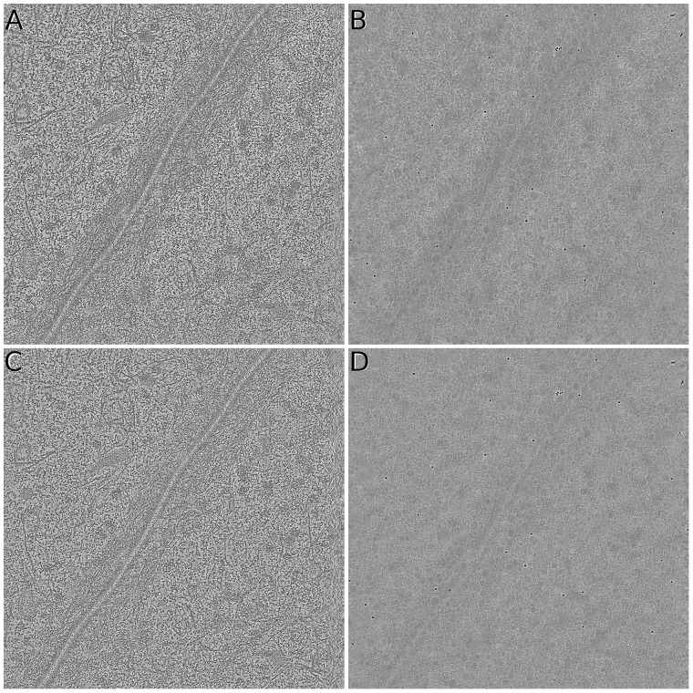
Motivation: Dual-axis electron tomography is an important 3 D macro-molecular structure reconstruction technology, which can reduce artifacts and suppress the effect of missing wedge. However, the fully automatic data process for dual-axis electron tomography still remains a challenge due to three difficulties: (i) how to track the mass of fiducial markers automatically; (ii) how to integrate the information from the two different tilt series; and (iii) how to cope with the inconsistency between the two different tilt series.

Results: Here we develop a toolkit for fully automatic alignment of dual-axis electron tomography, with a simultaneous reconstruction procedure. The proposed toolkit and its workflow carries out the following solutions: (i) fully automatic detection and tracking of fiducial markers under large-field datasets; (ii) automatic combination of two different tilt series and global calibration of projection parameters; and (iii) inconsistency correction based on distortion correction parameters and the consequently simultaneous reconstruction. With all of these features, the presented toolkit can achieve accurate alignment and reconstruction simultaneously and conveniently under a single global coordinate system.

Availability and implementation: The toolkit AuTom-dualx (alignment module dualxmauto and reconstruction module volrec_mltm) are accessible for general application at http://ear.ict.ac.cn, and the key source code is freely available under request.

Supplementary information: Supplementary data are available at Bioinformatics online.


Figures

Fig. 1.
Fig. 1.
The workflow of fully automatic simultaneous alignment scheme
Fig. 2.
Fig. 2.
The workflow of fiducial marker detection
Fig. 3.
Fig. 3.
Illustration of test datasets. (A)&(D) The 0° micrographs of Centriole of axis A and axis B. (B)&(E) The 0° micrographs of Adhesion belt of axis A and axis B. (C)&(F) The 0° micrographs of simulated dataset of axis A and axis B
Fig. 4.
Fig. 4.
Illustration of marker detection. All the detected markers are marked by circles. (A) Detected fiducial markers from the micrograph of axis A with 0° tilt angle. (B) Detected fiducial markers from the micrograph of axis A with 64° tilt angle. (C) Detected fiducial markers from the micrograph of axis B with 0° tilt angle. (D) Detected fiducial markers from the micrograph of axis B with 64° tilt angle
Fig. 5.
Fig. 5.
(A) The track lengths of tilt series of axis A. (B) The track lengths of tilt series of axis B. (C) The combined track lengths of tilt series of axis A and B
Fig. 6.
Fig. 6.
Illustration of pre-refinement of projection parameters. (A) Overlay of the pre-refined fiducial marker tracks of dataset Centriole. (B) Overlay of the pre-refined fiducial marker tracks of dataset Adhesion belt
Fig. 7.
Fig. 7.
Reconstruction results of the lefttop part of Centriole (SART with 40 iterations and relaxation factor λ=0.2). (A) Reconstruction of the alignment based on the classic orthogonal projection. (B) Reconstruction of the alignment with distortion correction
Fig. 8.
Fig. 8.
Sample slices of reconstruction on the Centriole dataset. All the illustrated data are reconstructed by SART with 40 iterations and relaxation factor λ=0.2. (A)&(B) The middle and top slices of the reconstruction obtained by the simultaneous alignment and reconstruction of AuTom-dualx. (C)&(D) The middle and top slices of the reconstruction obtained by the separate reconstruction and merging of IMOD (merging axis B to axis A)
Fig. 9.
Fig. 9.
Sample slices of reconstruction on the Adhesion belt dataset. All the illustrated data are reconstructed by SART with 40 iteration and relaxation factor λ=0.2. (A)&(B) The middle and top slices of the reconstruction obtained by the simultaneous alignment and reconstruction of AuTom-dualx. (C)&(D) are the middle and top slices of the reconstruction obtained by the separate reconstruction and merging of IMOD (merging axis B to axis A)
Fig. 10.
Fig. 10.
Fiducial marker subvolumes from the Centriole (left) and Adhesion belt (right) datasets. The fiducial marker location in the Centriole volumes is (449 418 259) for AuTom-dualx and (475 418 283) for IMOD. The fiducial marker location in the Adhesion belt volumes is (578 1025 116) for AuTom-dualx and (574 1035 105) for IMOD
Fig. 11.
Fig. 11.
The NCC curves between the reprojection of the reconstruction for both AuTom-dualx and IMOD and the original tilt series for the Centriole dataset (A) and the Adhesion belt dataset (B)
Fig. 12.
Fig. 12.
The Fourier shell correlation between the reconstructed subtomogram and the reference 2WRJ map
Fig. 13.
Fig. 13.
Results obtained by Resmap analysis. (A)&(C)&(E) The slices, local resolution distribution and resolution histogram of the subtomogram produced by the workflow of AuTom-dualx, respectively. (B)&(D)&(F) The slices, local resolution distribution and resolution histogram of the subtomogram produced by the workflow of IMOD, respectively

References

    1. Aiger D., et al. (2008) 4-points congruent sets for robust pairwise surface registration. ACM Trans. Graphic, 27, 85.
    1. Andersen A.H., Kak A.C. (1984) Simultaneous algebraic reconstruction technique (sart): a superior implementation of the art algorithm. Ultrason. Imaging, 6, 81–94. - PubMed
    1. Arslan I., et al. (2006) Reducing the missing wedge: high-resolution dual axis tomography of inorganic materials. Ultramicroscopy, 106, 994–1000. - PubMed
    1. Bharat T.A., Scheres S.H. (2016) Resolving macromolecular structures from electron cryo-tomography data using subtomogram averaging in relion. Nat. Protoc., 11, 2054. - PMC - PubMed
    1. Cantele F., et al. (2007) Local refinement: an attempt to correct for shrinkage and distortion in electron tomography. J. Struct. Biol., 158, 59–70. - PMC - PubMed

Publication types