

Programmed Death Ligand 1 Is a Negative Prognostic Marker in Recurrent Isocitrate Dehydrogenase-Wildtype Glioblastoma
- PMID: 30011045
- PMCID: PMC7137460
- DOI: 10.1093/neuros/nyy268
Programmed Death Ligand 1 Is a Negative Prognostic Marker in Recurrent Isocitrate Dehydrogenase-Wildtype Glioblastoma
Abstract
Background: Checkpoint inhibition has demonstrated clinical efficacy in a variety of solid tumors. Reports of programmed death ligand 1 (PD-L1) expression in glioblastoma are highly variable (ranging from 6% to 88%) and its role as a prognostic marker has yielded conflicting results.
Objective: To validate the prevalence and prognostic role of PD-L1 expression in a large cohort of diffuse gliomas according to the 2016 revised WHO classification.
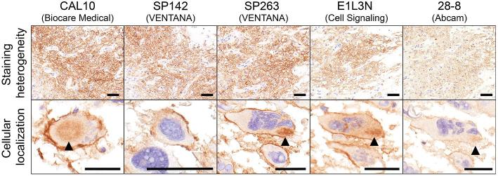
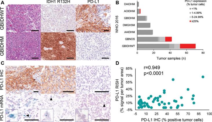
Methods: Using tissue microarrays, we compared 5 PD-L1 monoclonal antibodies (n = 56) and validated expression (n = 183) using quantitative immunohistochemistry (IHC) and RNA in situ hybridization (RISH). Expression data from The Cancer Genome Atlas (TCGA) and published studies were compared with clinical outcome. Multiplexed immunophenotyping was used to identify PD-L1+ cell populations in post-treatment glioblastoma.
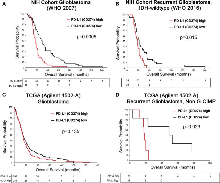
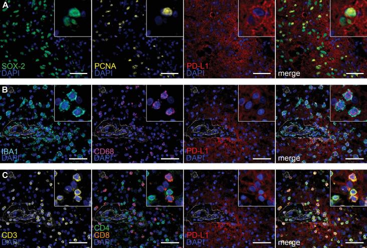
Results: Using a 5% cut-off, PD-L1 expression was significantly associated with a poor prognosis in both histologically defined (n = 125, log-rank P < .001) and recurrent isocitrate dehydrogenase (IDH)-wildtype glioblastoma (n = 60, log-rank P = .015). PD-L1 remained a significant negative prognosticator in Cox regression analysis (hazard ratio: 1.96, P = .021). Analysis of TCGA data confirmed decreased overall survival in recurrent non-glioma CpG island methylator phenotype (G-CIMP) glioblastoma (n = 12, log-rank P = .023), but not in glioblastoma as a group (n = 444, log-rank P = .135). PD-L1 RISH showed a significant correlation with IHC (P < .0001). PD-L1 was observed in the proliferating perivascular stem cell and immune niche of post-treatment glioblastoma.
Conclusion: A 5% PD-L1 expression cut-off identified a subset of glioblastoma that is associated with a worse clinical outcome. This association remained significant within the newly defined IDH-wildtype classification. These findings could have implications for patient stratification in future clinical trials of PD-1/PD-L1 blockade.
Keywords: Glioblastoma; Immunohistochemistry; Immunotherapy; In situ hybridization; PD-1; PD-L1; RNA.
Published by Oxford University Press on behalf of Congress of Neurological Surgeons 2018.
Figures
References
-
- Dong H, Strome SE, Salomao DR, et al.. Erratum: Tumor-associated B7-H1 promotes T-cell apoptosis: a potential mechanism of immune evasion. Nat Med. 2002;8(8):793-800. - PubMed
-
- Wintterle S, Schreiner B, Mitsdoerffer M, et al.. Expression of the B7-related molecule B7-H1 by glioma cells: a potential mechanism of immune paralysis. Cancer Res. 2003;63(21):7462-7467. - PubMed
MeSH terms
Substances
LinkOut - more resources
Full Text Sources
Other Literature Sources
Medical
Research Materials
Miscellaneous
