Practice Guideline
. 2018 Aug;156(2):e41-e74.
doi: 10.1016/j.jtcvs.2018.02.115.

The American Association for Thoracic Surgery consensus guidelines on bicuspid aortic valve-related aortopathy: Full online-only version

Affiliations
Practice Guideline

The American Association for Thoracic Surgery consensus guidelines on bicuspid aortic valve-related aortopathy: Full online-only version

Michael A Borger et al. J Thorac Cardiovasc Surg. 2018 Aug.

Abstract

Bicuspid aortic valve disease is the most common congenital cardiac disorder, being present in 1% to 2% of the general population. Associated aortopathy is a common finding in patients with bicuspid aortic valve disease, with thoracic aortic dilation noted in approximately 40% of patients in referral centers. Several previous consensus statements and guidelines have addressed the management of bicuspid aortic valve-associated aortopathy, but none focused entirely on this disease process. The current guidelines cover all major aspects of bicuspid aortic valve aortopathy, including natural history, phenotypic expression, histology and molecular pathomechanisms, imaging, indications for surgery, surveillance, and follow-up, and recommendations for future research. It is intended to provide clinicians with a current and comprehensive review of bicuspid aortic valve aortopathy and to guide the daily management of these complex patients.


Conflict of interest statement

Conflict of Interest Statement

Dr Borger received speakers’ honoraria and/or consulting fees from Edwards Lifesciences, Medtronic, St Jude Medical, and CryoLife. Dr Moon received speakers’ honoraria and/or consulting fees from Edwards Lifesciences and Medtronic. All other authors have nothing to disclose with regard to commercial support.

Figures

FIGURE 1.
FIGURE 1.
Sievers’ classification system for BAVas viewed from the surgeon’s side with the left coronary artery at left. The number of specimens is given, and the percentage is shown in parentheses. The blackened lines represent raphe. The main category is based on the number of raphes, the first subcategory is based on spatial position, and the second subcategory reflects valve function. Ap, Anterior-posterior; B, balanced valvular lesion; I, insufficiency; L, left coronary sinus; lat, lateral; N, noncoronary sinus; No, normal function; R, right coronary sinus; S, stenosis. Used with permission from Sievers and Schmidtke.
FIGURE 2.
FIGURE 2.
Anatomic distribution of aortic aneurysms in patients with BAV. BAV, Bicuspid aortic valve. (Aortic Institute at Yale-New Haven, unpublished data, June 30, 2017.) These data are similar to those reported by Michelena and colleagues.
FIGURE 3.
FIGURE 3.
A, Normal aortic valve anatomy with an opening angle of 75 degrees, flow jet angle (θ1, which measures displacement of peak systolic flow [arrow] from vessel midline). B, BAV showing restricted valve opening (opening angle of 60 degrees), displaced flow jet with associated θ1, displaced high-flow velocities near the vessel wall leading to asymmetrically increased WSS at the aortic convexity. WSS, Wall shear stress. Reproduced with permission from Burris and Hope.
FIGURE 4.
FIGURE 4.
Root phenotype versus ascending phenotype of BAV aortopathy. Echocardiographic imaging is shown in the upper left corner, 3-dimensional reconstruction is shown in lower left corner, and intraoperative findings are shown at right. In the root phenotype, the diameter of the aorta at the level of the sinuses of Valsalva is greater than that of the tubular ascending aorta, whereas in the ascending phenotype the diameter of the tubular ascending aorta is greater than that of the sinuses.
FIGURE 5.
FIGURE 5.
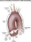
Subdivision of proximal aortic involvement in BAV aortopathy. Reproduced with permission from Verma and Siu.
FIGURE 6.
FIGURE 6.
Typical echocardiographic findings in a patient with BAV with tubular ascending aorta dilatation phenotype. A, Echocardiogram of a 60-year-old woman with R/N BAV, no aortic valve regurgitation, and a fusiform ascending tubular aortic aneurysm. Left parasternal long-axis view in diastole shows root measurement of 36 mm (first arrow from left) and midtubular ascending aorta measurement of 47 mm (second arrow from left). B, Suprasternal diastolic view shows the mildly dilated proximal arch (36 mm, arrow) and normal upper descending aorta. C, Parasternal short-axis en face view of the aortic valve in systole shows 2 commissures (asterisks) at 1 and 7 o’clock with right nonfusion. LV, Left ventricle; RV, right ventricle; Ao, aorta; PA, pulmonary artery; RA, right atrium; LA, left atrium.
FIGURE 7.
FIGURE 7.
Patient with BAV with root phenotype aortic dilation. A, Echocardiogram of a 53-year-old man with R/L BAV, severe aortic valve regurgitation, and root-proximal ascending aortic aneurysm. Left parasternal long-axis view in diastole shows root measurement of 46 mm (arrow), STJ effacement (asterisk), and proximal tubular ascending aorta dilatation. B, Left parasternal long-axis zoomed color-Doppler view in diastole shows the flow convergence (arrow) of a posteriorly directed jet that quantified to 78 mL per beat of regurgitant volume. C, Parasternal short-axis en face view of the aortic valve in systole shows 2 commissures (asterisks) at 4 and 10 o’clock with right-left fusion. LV, Left ventricle; RV, right ventricle; LA, left atrium; Ao, aorta; RA, right atrium.
FIGURE 8.
FIGURE 8.
MRI assessment of patients with BAV showing normal aortas (left) and different types of BAV aortopathy (middle and right). Each of the 3 upper panels shows maximum intensity projection of magnetic resonance angiography with the corresponding inferior panel demonstrating the planar analysis of systolic flow. The left panel demonstrates imaging from a normal patient. The middle panel demonstrates aneurysmal dilation at the level of the sinuses with flow directed rightward and posteriorly in a patient with a left-right cusp fusion. The right panel shows more diffuse aneurysmal dilation in a patient with right-noncoronary cusp fusion and flow directed leftward and posteriorly. RA, Right anterior; RP, right posterior; LA, left anterior; LP, left posterior. Adapted from Burris and Hope.
FIGURE 9.
FIGURE 9.
CT imaging with 3 dimensional reconstruction of a patient with BAV with associated aortopathy.
FIGURE 10.
FIGURE 10.
Transthoracic assessment for aortic coarctation. A, Echocardiogram of a 31-year-old woman with BAV and severe aortic coarctation. Suprasternal systolic still frame shows laminar Doppler flow through the proximal portion of the arch (“ARCH”) before becoming turbulent flow across a tight coarctation (arrow) just distal to the left subclavian (asterisk). B, Suprasternal diastolic still frame shows no Doppler flow through the proximal portion of the arch but persistent diastolic turbulent flow across the coarctation (arrow) just distal to the left subclavian (asterisk). C, Continuous-wave Doppler signal across the coarctation shows a systolic (measurement) peak gradient of 64 mm Hg through the coarctation, with persistent flow in diastole (arrow). D, Pulsed-wave Doppler signal of the abdominal aorta shows a delayed peaking of the systolic signal (line) with prominent persistent flow in diastole (arrow), pathognomonic of coarctation.
FIGURE 11.
FIGURE 11.
A, Schematic shows the leading-edge to leading-edge measurement technique used in echocardiography, from left to right: measurement of the sinuses of Valsalva, STJ, and proximal tubular ascending aorta. B, Inner-to-inner measurements used in MRI and CT. In addition, a consistent approach to measuring all 3 sinuses with MRI and CT is necessary. The sinus-to-commissure and sinus-to-sinus measurements can both be used, but consistency is necessary for interval surveillance. C, Standard measurement locations for MRI and CTwith the inner-wall to inner-wall technique. RCA, Right coronary artery; LCA, left coronary artery; MRI, magnetic resonance imaging; CT, computed tomography; DAo, descending aorta.
FIGURE 12.
FIGURE 12.
TEE aorta assessment. A, Prebypass echocardiogram of a 79-year-old man with typical BAV (right-left cusp fusion), mild AS, and severe generalized aorta dilatation. High-esophageal mid-ascending aorta short-axis measurement (arrow) at 0°. B, Same imaging position as A, now at 91°, reveals the mid-ascending aorta at 52 mm (long arrow) and the distal aorta (short arrow) at 49 mm. C, Mid-esophageal long axis at 127° allows measurements of the proximal ascending aorta and root (dotted lines). D, Mid-esophageal long axis at 139° allows improved visualization of the root, which measured 49 mm (dotted line). The patient underwent a Bentall procedure. Ao, Aorta; RPA, right pulmonary artery; RV, right ventricle; LV, left ventricle.
FIGURE 13.
FIGURE 13.
A, 2D balanced steady-state free precession cine MRI showing the left ventricular outflow tract and the position of the aortic valve imaging plane shown in yellow. B, TAV and the 2 most common BAV phenotypes: R/L and R/N cusp fusion. Arrows show the location of the raphe (if present) between the conjoined cusps. The conjoined R/L cusp (yellow box, arrow) is also seen to be doming in the corresponding left ventricular outflow tract view (A, arrow). Bicuspidality of the aortic valve should be assessed in systole rather than diastole, because the valves often appear tricuspid when closed. TAV, Tricuspid aortic valve; RL, right-left; RN, right noncoronary. Adapted from Entezari and colleagues.
FIGURE 14.
FIGURE 14.
MRI of a 73-year-old man shows the (A) balanced steady-state free precession valve cines of a patient with BAV with R/L fusion and no stenosis. B, Contrast-enhanced magnetic resonance angiography shows mild dilation of the sinus of Valsalva with a maximal dimension of 40 mm and (C) a 47-mm dilation of the mid-ascending aorta. D, An eccentric jet is observed downstream from the nonstenotic bicuspid valve that impacts along the anterior portion of the tubular aorta. L, Left coronary cusp; N, noncoronary cusp; R, coronary cusp.

References

    1. Hoffman J, Kaplan S. The incidence of congenital heart disease. J Am Coll Cardiol 2002;39:1890–900. - PubMed
    1. Masri A, Kalahasti V, Alkharabsheh S, Svensson LG, Sabik JF, Roselli EE, et al. Characteristics and long-term outcomes of contemporary patients with bicuspid aortic valves. J Thorac Cardiovasc Surg 2016;151:1650–1659.e1. - PubMed
    1. Januzzi JL, Isselbacher EM, Fattori R, Cooper JV, Smith DE, Fang J, et al. Characterizing the young patient with aortic dissection: results from the International Registry of Aortic Dissection (IRAD). J Am Coll Cardiol 2004;43:665–9. - PubMed
    1. Fedak P, David T, Borger M, Verma S, Butany J, Weisel R. Bicuspid aortic valve disease: recent insights in pathophysiology and treatment. Expert Rev Cardiovasc Ther 2005;3:295–308. - PubMed
    1. Hiratzka LF, Bakris GL, Beckman JA, Bersin RM, Carr VF, Casey DE Jr, et al. 2010 ACCF/AHA/AATS/ACR/ASA/SCA/SCAI/SIR/STS/SVM guidelines for the diagnosis and management of patients with Thoracic Aortic Disease: a report of the American College of Cardiology Foundation/American Heart Association Task Force on practice guidelines, American Association for Thoracic Surgery, American College of Radiology, American Stroke Association, Society of Cardiovascular Anesthesiologists, Society for Cardiovascular Angiography and Interventions, Society of Interventional Radiology, Society of Thoracic Surgeons, and Society for Vascular Medicine. Circulation 2010; 121:e266–369. - PubMed

Publication types

MeSH terms