Cranial neural tube defect after trimethoprim exposure
- PMID: 30012199
- PMCID: PMC6048906
- DOI: 10.1186/s13104-018-3593-1
Cranial neural tube defect after trimethoprim exposure
Abstract
Objectives: The Neural Tube Defects Research Group of University of Malaya was approached to analyze a tablet named TELSE, which may have resulted in a baby born with central nervous system malformation at the University of Malaya Medical Centre. In this animal experimental study, we investigated the content of TELSE and exposure of its contents that resulted in failure of primary neurulation.
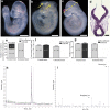
Results: Liquid Chromatography Tandem Mass spectrophotometry analysis of the TELSE tablet confirmed the presence of trimethoprim as the active compound. The TELSE tablet-treated females produced significant numbers of embryos with exencephaly (n = 8, 36.4%, *P < 0.0001), in all litters. The TELSE tablet-treated females subsequently given folic acid did not result in pregnancies despite there being evidence of possible resorption. Furthermore, after multiple rounds of mating which did not yield viable pregnancies, eventually, 2 embryos with exencephaly were harvested in a litter of 6 at 0.05% w/v pure trimethoprim once. The use of trimethoprim, a folic acid antagonist, peri-conceptionally increased the risk of exencephaly in the mouse.
Keywords: Acne; Folic acid antagonist; Malaysia; Neural tube defects; Primary neurulation; Trimethoprim.
Figures

References
-
- Sahmat A, Gunasekaran R, Mohd-Zin SW, Balachandran L, Thong MK, Engkasan JP, Ganesan D, Omar Z, Azizi AB, Ahmad-Annuar A, Abdul-Aziz NM. The prevalence and distribution of spina bifida in a single major referral center in Malaysia. Front Pediatr. 2017; 5:237. 10.3389/fped.2017.00237. eCollection 2017. - PMC - PubMed
MeSH terms
Substances
Grants and funding
- UM.C/625/1/HIR/062/University of Malaya High Impact Research Grant
- UM.C/625/1/HIR/062/University of Malaya High Impact Research Grant
- UM.C/625/1/HIR/062/University of Malaya High Impact Research Grant
- UM.C/625/1/HIR/062/University of Malaya High Impact Research Grant
- UM.C/625/1/HIR/148/2/University of Malaya High Impact Research Grant
- UM.C/625/1/HIR/148/2/University of Malaya High Impact Research Grant
- UM.C/625/1/HIR/148/2/University of Malaya High Impact Research Grant
- UM.C/625/1/HIR/148/2/University of Malaya High Impact Research Grant
- UM.C/625/1/HIR/MOHE/MED/08/Ministry of Higher Education University of Malaya High Impact Research Grant
- UM.C/625/1/HIR/MOHE/MED/08/Ministry of Higher Education University of Malaya High Impact Research Grant
- UM.C/625/1/HIR/MOHE/MED/08/Ministry of Higher Education University of Malaya High Impact Research Grant
- UM.C/625/1/HIR/MOHE/MED/08/Ministry of Higher Education University of Malaya High Impact Research Grant
- PG153-2015B/University of Malaya Postgraduate Research Grant (PPP)
- PG153-2015B/University of Malaya Postgraduate Research Grant (PPP)
- PG137-2015A/University of Malaya Postgraduate Research Grant (PPP)
- PG137-2015A/University of Malaya Postgraduate Research Grant (PPP)
LinkOut - more resources
Full Text Sources
Other Literature Sources
Medical

