

Recessive mutations in ATP8A2 cause severe hypotonia, cognitive impairment, hyperkinetic movement disorders and progressive optic atrophy
- PMID: 30012219
- PMCID: PMC6048855
- DOI: 10.1186/s13023-018-0825-3
Recessive mutations in ATP8A2 cause severe hypotonia, cognitive impairment, hyperkinetic movement disorders and progressive optic atrophy
Abstract
Background: ATP8A2 mutations have recently been described in several patients with severe, early-onset hypotonia and cognitive impairment. The aim of our study was to characterize the clinical phenotype of patients with ATP8A2 mutations.
Methods: An observational study was conducted at multiple diagnostic centres. Clinical data is presented from 9 unreported and 2 previously reported patients with ATP8A2 mutations. We compare their features with 3 additional patients that have been previously reported in the medical literature.
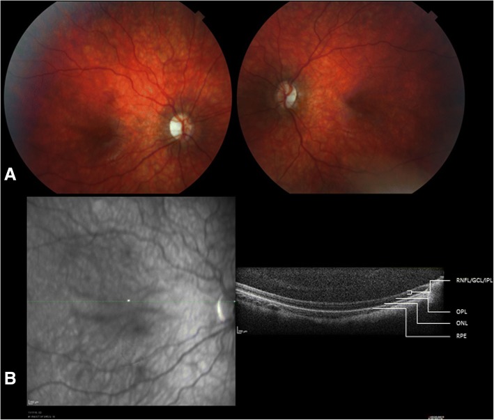
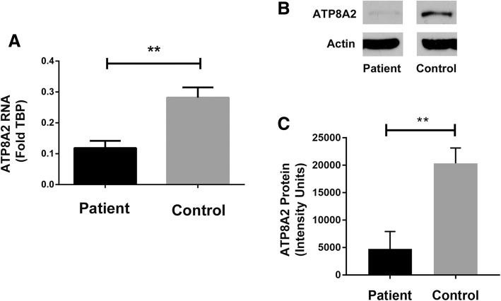
Results: Eleven patients with biallelic ATP8A2 mutations were identified, with a mean age of 9.4 years (range 2.5-28 years). All patients with ATP8A2 mutations (100%) demonstrated developmental delay, severe hypotonia and movement disorders, specifically chorea or choreoathetosis (100%), dystonia (27%) and facial dyskinesia (18%). Optic atrophy was observed in 78% of patients for whom funduscopic examination was performed. Symptom onset in all (100%) was noted before 6 months of age, with 70% having symptoms noted at birth. Feeding difficulties were common (91%) although most patients were able to tolerate pureed or thickened feeds, and 3 patients required gastrostomy tube insertion. MRI of the brain was normal in 50% of the patients. A smaller proportion was noted to have mild cortical atrophy (30%), delayed myelination (20%) and/or hypoplastic optic nerves (20%). Functional studies were performed on differentiated induced pluripotent cells from one child, which confirmed a decrease in ATP8A2 expression compared to control cells.
Conclusions: ATP8A2 gene mutations have emerged as the cause of a novel neurological phenotype characterized by global developmental delays, severe hypotonia and hyperkinetic movement disorders, the latter being an important distinguishing feature. Optic atrophy is common and may only become apparent in the first few years of life, necessitating repeat ophthalmologic evaluation in older children. Early recognition of the cardinal features of this condition will facilitate diagnosis of this complex neurologic disorder.
Keywords: ATP8A2; Chorea; Choreoathetosis; Developmental disabilities; Dystonia; Optic atrophy; Phospholipid transfer protein; Whole exome sequencing.
Conflict of interest statement
Ethics approval and consent to participate
The Research Ethics Board of the Hospital for Sick Children approved this study and informed consent was obtained from all families included in this study.
Consent for publication
All parents provided written informed consent for the publication of the videos used in this report.
Competing interests
The authors declare that they have no competing interests.
Publisher’s Note
Springer Nature remains neutral with regard to jurisdictional claims in published maps and institutional affiliations.
Figures
References
Publication types
MeSH terms
Substances
Grants and funding
LinkOut - more resources
Full Text Sources
Other Literature Sources
Molecular Biology Databases
