. 2018 Jul 31;115(31):E7265-E7274.
doi: 10.1073/pnas.1803916115. Epub 2018 Jul 16.

Pupil mimicry promotes trust through the theory-of-mind network

Affiliations

Pupil mimicry promotes trust through the theory-of-mind network

Eliska Prochazkova et al. Proc Natl Acad Sci U S A. .

Abstract

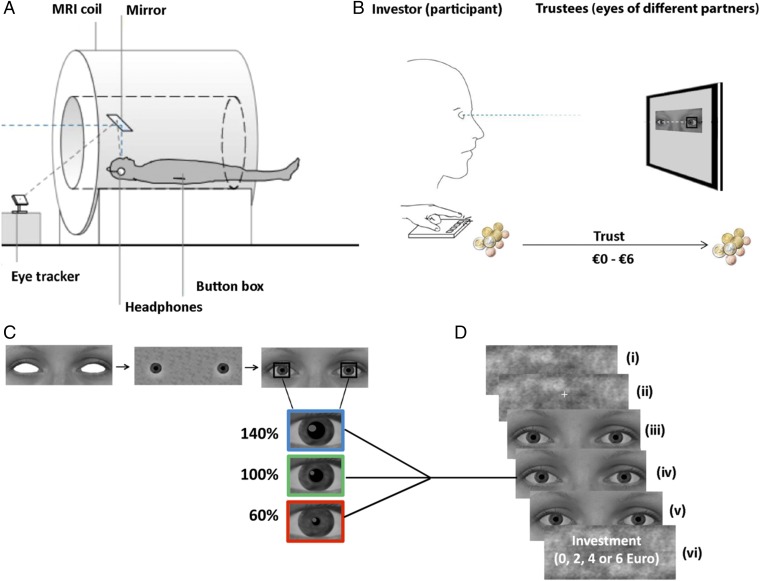
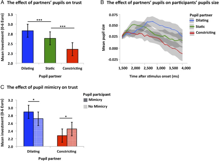
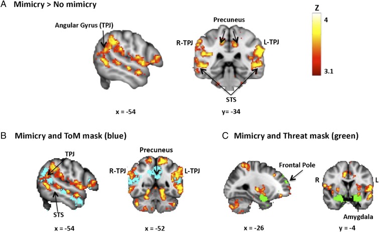
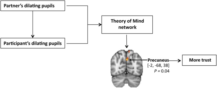
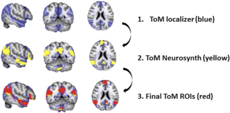
The human eye can provide powerful insights into the emotions and intentions of others; however, how pupillary changes influence observers' behavior remains largely unknown. The present fMRI-pupillometry study revealed that when the pupils of interacting partners synchronously dilate, trust is promoted, which suggests that pupil mimicry affiliates people. Here we provide evidence that pupil mimicry modulates trust decisions through the activation of the theory-of-mind network (precuneus, temporo-parietal junction, superior temporal sulcus, and medial prefrontal cortex). This network was recruited during pupil-dilation mimicry compared with interactions without mimicry or compared with pupil-constriction mimicry. Furthermore, the level of theory-of-mind engagement was proportional to individual's susceptibility to pupil-dilation mimicry. These data reveal a fundamental mechanism by which an individual's pupils trigger neurophysiological responses within an observer: when interacting partners synchronously dilate their pupils, humans come to feel reflections of the inner states of others, which fosters trust formation.

Keywords: affect; neuroimaging; physiological linkage; social cognition; trust game.


Conflict of interest statement

The authors declare no conflict of interest.

Figures

Fig. 1.
Fig. 1.
Experimental set-up, stimuli, and task. (A) Inside the MRI scanner, the participants played one-player trust-games while their investment-decisions and pupil diameter were measured with a button box and eye-tracker, respectively. (B) Subjects (investors) watched short video clips showing the eye region of different virtual partners (trustees) whose pupils were manipulated to change in size. In each trial, subjects were asked to transfer between €0 and €6 to their partner. Investments were then tripled and the virtual trustee was asked to transfer between 0% and 100% of the tripled amount back to the investor. No feedback was provided so that subject’s investments (indicating trust) were based on information from the partner’s eye region only. (C) The stimulus material consisted of 18 photos with neutral expressions (nine males). The eyes were then filled with eye whites and irises, and an artificial pupil was added. The partner’s pupil dilated (140% of the original diameter), constricted (60%), or remained static (range of 3–7 mm). (D) Stimuli presentation. (i) A Fourierscrambled image was presented for 4,000 ms; (ii) fixation followed for 500 ms; (iii) the eye stimulus remained static for the first 1,500 ms; then (iv) in the dilation and constriction conditions, the pupils gradually changed in size over 1,500 ms; and (v) remained static at that size during the final 1,000 ms (in the static condition, pupils remained at the same size throughout the trial). (vi) A screen appeared asking participants to make an investment decision.
Fig. 2.
Fig. 2.
Behavioral and pupillometry results. (A) The bar plot shows that mean trust-related investments (€) increased in response to partners’ dilating pupil size (n = 40 participants). Error bars indicate ±1 SE. ***P < 0.001 for each factor, pairwise contrasts: dilating pupils vs. static pupils [B = 0.19, CI (0.08, 0.30)] and constricting pupils vs. static pupils [B = −0.28, CI (−0.38, −0.17)]. (B) Participants mimicked partner’s pupil sizes: the curves correspond to participants’ mean pupil response from baseline over the remaining of stimulus presentation time (ms), in response to partner’s dilating, static, and constricting pupils. Mean pupil size is depicted in arbitrary values. Shaded areas indicate the 99% CI. (C) The bar plot shows mean investments (€) as a function of partners’ and participants’ pupil size. Error bars indicate ±1 SE. *P < 0.01. Mean investment increases when a participant’s own pupils dilate in response to their partner’s dilating pupils. Pairwise contrast: pupil-dilation mimicry vs. no pupil-dilation mimicry [B = 0.175, CI (0.02, 0.33)]. Mean investment decreases when participants’ pupil constricts in response to their partners’ constricting pupils. Pairwise contrast: pupil-constriction mimicry vs. no pupil-constriction mimicry [B = −0.173, CI (−0.33, −0.02)].
Fig. 3.
Fig. 3.
Neural correlates of pupil mimicry. (A) During mimicry, subjects displayed enhanced ToM activation. Peak voxels MNI x, y, z coordinates TPJ [60, −54, 18/−58, −54, 18], bilateral STS [52, −34, 2/−52, −34, 2], right MPFC [6, 46, 8] (not displayed in the image), precuneus cortex [8, −40, 48/−8, −40, 48]; threshold at P < 0.05 [cluster-level FWE correction with multiple comparisons at 2.3, (n = 34 participants)]. For visualization, the threshold was set at z = 3.1–4. (B and C) The image shows additional overlaps between pupil-mimicry pattern and ToM network (blue) and threat network (green). The background images reflect MNI 2-mm template (0.05-voxel size smoothing kernel); the right side of the image corresponds to the left side of the brain. Location coordinates are in stereotactic MNI space with a 2 × 2 × 2-voxel size. The source of anatomical labels is FSL Atlas tools.
Fig. 4.
Fig. 4.
ROI analyses. (A) Example of one subject’s ToM network mask selection. The ROIs were classified by an independent TOM localizer (blue) as well as by the additional inclusion of ToM masks (yellow), derived from our metaanalyses on previous studies. The overlapping voxels were used as the final mask (red). (B) The bar plot displays the mean parameter estimates averaged across all subjects (n = 33 participants) of the neural activation extracted from the individual ToM masks during four experimental conditions. During pupil-dilation mimicry the ToM percentage signal increase was significantly greater compared with all of the other conditions, including pupil-constriction mimicry [mean difference = −0.098, SE = 0.03, CI (0.02, 0.18), P = 0.005], no-constricting mimicry [mean difference = −0.113, SE = 0.03, CI (0.03, 0.19), P = 0.005], and no-dilation mimicry conditions [mean difference = −0.116, SE = 0.02, CI (0.05, 0.18), P < 0.001]. **P < 0.01, ***P < 0.001. Error bars indicate ±1 SE. (C) The scatter plot shows that the same subjects that displayed larger pupil-dilation mimicry index also displayed the greatest increase in ToM percentage signal during pupil-dilation mimicry (R = 0.47, P < 0.01).
Fig. 5.
Fig. 5.
ToM and trust. The diagram shows that the ToM network is modulated by pupil-dilation mimicry. Within the ToM network, investment rates predicted precuneus BOLD signal changes, confirming ToM involvement in trust decisions. Peak voxel MNI x, y, z coordinates [−2, −68, 38], corrected for ToM network with threshold free cluster enhancement (42) (threshold z = 3.1, P < 0.05, FWE-corrected P value from the minimum voxels in the cluster).
Fig. 6.
Fig. 6.
ROIs selection example from one subject. The final ToM mask included MNI coordinates mentioned by Saxe and Kanwisher (63). These were [−54, −60, 21] for the left TPJ, [51, −54, 27] for the right TPJ, [−9, −51, 33] for the precuneus, [−57, −27, −12] for the left anterior STS, and [66, −18, −15] for the right anterior STS. All subjects shared activation in threat ROIs: amygdala [24, 2, −20/−22, 0, −22], frontal pole: [−24, 58, 16], brainstem [2, −24, −14]. The background image reflects MNI 2-mm template (0.05 voxel size smoothing kernel).

Comment in

References

    1. Fehr E, Gächter S. Altruistic punishment in humans. Nature. 2002;415:137–140. - PubMed
    1. Rand DG, Greene JD, Nowak MA. Spontaneous giving and calculated greed. Nature. 2012;489:427–430. - PubMed
    1. Tamietto M, de Gelder B. Neural bases of the non-conscious perception of emotional signals. Nat Rev Neurosci. 2010;11:697–709. - PubMed
    1. De Dreu CKW, et al. In-group defense, out-group aggression, and coordination failures in intergroup conflict. Proc Natl Acad Sci USA. 2016;113:10524–10529. - PMC - PubMed
    1. Hess EH. The role of pupil size in communication. Sci Am. 1975;233:110–112, 116–119. - PubMed

Publication types

LinkOut - more resources