. 2018 Jul;11(7):e002038.
doi: 10.1161/CIRCGEN.117.002038.

Multigenic Disease and Bilineal Inheritance in Dilated Cardiomyopathy Is Illustrated in Nonsegregating LMNA Pedigrees

Affiliations

Multigenic Disease and Bilineal Inheritance in Dilated Cardiomyopathy Is Illustrated in Nonsegregating LMNA Pedigrees

Jason R Cowan et al. Circ Genom Precis Med. 2018 Jul.

Abstract

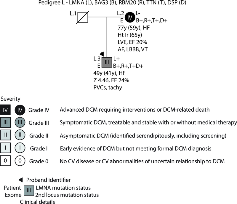
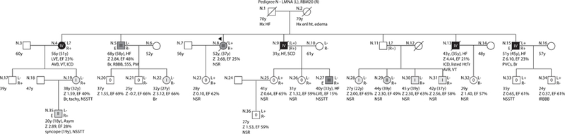
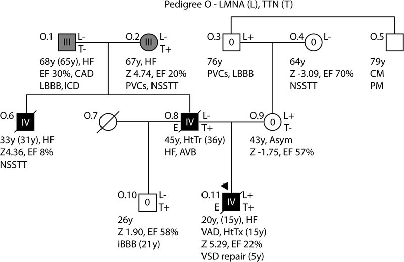
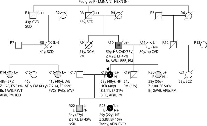
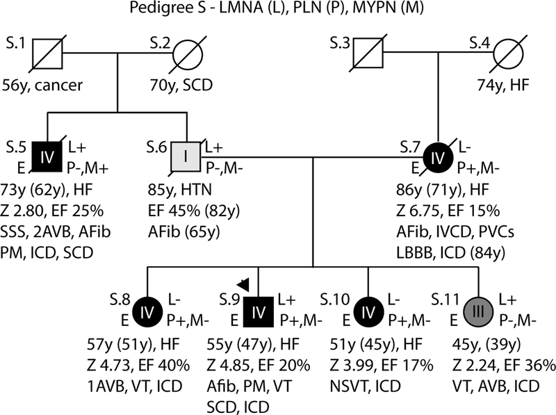
Background: We have previously described 19 pedigrees with apparent lamin (LMNA)-related dilated cardiomyopathy (DCM) manifesting in affected family members across multiple generations. In 6 of 19 families, at least 1 individual with idiopathic DCM did not carry the family's LMNA variant. We hypothesized that additional genetic cause may underlie DCM in these families.

Methods: Affected family members underwent exome sequencing to identify additional genetic cause of DCM in the 6 families with nonsegregating LMNA variants.

Results: In 5 of 6 pedigrees, we identified at least 1 additional rare variant in a known DCM gene that could plausibly contribute to disease in the LMNA variant-negative individuals. Bilineal inheritance was clear or presumed to be present in 3 of 5 families and was possible in the remaining 2. At least 1 individual with a LMNA variant also carried a variant in an additional identified DCM gene in each family. Using a multivariate linear mixed model for quantitative traits, we demonstrated that the presence of these additional variants was associated with a more severe phenotype after adjusting for sex, age, and the presence/absence of the family's nonsegregating LMNA variant.

Conclusions: Our data support DCM as a genetically heterogeneous disease with, at times, multigene causation. Although the frequency of DCM resulting from multigenic cause is uncertain, our data suggest it may be higher than previously anticipated.

Keywords: cardiomyopathy, dilated; exome sequencing; genetics; inheritance patterns; lamin type A; multifactorial inheritance; pedigree.


Figures

Figure 1:
Figure 1:. Pedigrees with non-segregating LMNA variants, Pedigree L.
As per convention, the proband for each family is indicated by an arrow. Males are shown as squares and females as circles. Deceased individuals are denoted by a diagonal slash. Individuals for whom exome sequencing was completed are marked on the left side with an E. Genetic testing results are indicated on the right side by either a + (variant present) or a – (variant absent). Gene abbreviations are provided in the title of each pedigree. Obligate carriers are marked in parentheses (+). The upper right symbol reports genetic testing results for LMNA, while the bottom right symbols report testing results for additional identified loci (Table 2). Clinical data is structured as previously described. First line: current age if the individual is still living or age of death if the individual is deceased, age of onset (in parentheses), major diagnosis or cause of death. Second line: age of transplantation, if applicable. Third line: left ventricular size defined by magnitude of standard deviation (Z-score, z) and function defined by left ventricular ejection fraction (EF, %). Fourth line: arrhythmias or other conduction system disease. Abbreviations: AFib, atrial fibrillation; Asym, asymptomatic; AVB atrioventricular block; 1AVB or 2AVB, first-degree or second-degree AVB, respectively; A/W, alive and well; BiFB, bifascicular block; Br, bradycardia; CAD, coronary artery disease; CHD, congenital heart disease; CM, cardiomyopathy, unknown type; CVD, cardiovascular disease; DCM, dilated cardiomyopathy; EF, ejection fraction; HF, heart failure; HTN, hypertension; HtTr, heart transplant; ICD, implantable cardioverter defibrillator; IDC, idiopathic dilated cardiomyopathy; IBBB, incomplete bundle branch block; IRBBB or ILBBB, right or left IBBB, respectively; IVCD, interventricular conduction delay; LBBB, left bundle branch block; MVP, mitral valve prolapse; NSR, normal sinus rhythm; NSSTT, nonspecific ST-T changes on ECG; NSVT, non-sustained ventricular tachycardia; PACs, paroxysmal atrial contractions; PM, pacemaker; PSVT, paroxysmal supraventricular tachycardia; PVCs, premature ventricular contractions; RBBB, right bundle branch block; SCD, sudden cardiac death; tachy, sinus tachycardia; SSS, sick sinus syndrome; VSD, ventricular septal defect; VT, ventricular tachycardia; Z, Z-score of left ventricular end-diastolic dimension.
Figure 2:
Figure 2:. Pedigrees with non-segregating LMNA variants, Pedigree N.
As per convention, the proband for each family is indicated by an arrow. Males are shown as squares and females as circles. Deceased individuals are denoted by a diagonal slash. Individuals for whom exome sequencing was completed are marked on the left side with an E. Genetic testing results are indicated on the right side by either a + (variant present) or a – (variant absent). Gene abbreviations are provided in the title of each pedigree. Obligate carriers are marked in parentheses (+). The upper right symbol reports genetic testing results for LMNA, while the bottom right symbols report testing results for additional identified loci (Table 2). Clinical data is structured as previously described. First line: current age if the individual is still living or age of death if the individual is deceased, age of onset (in parentheses), major diagnosis or cause of death. Second line: age of transplantation, if applicable. Third line: left ventricular size defined by magnitude of standard deviation (Z-score, z) and function defined by left ventricular ejection fraction (EF, %). Fourth line: arrhythmias or other conduction system disease. Abbreviations: AFib, atrial fibrillation; Asym, asymptomatic; AVB atrioventricular block; 1AVB or 2AVB, first-degree or second-degree AVB, respectively; A/W, alive and well; BiFB, bifascicular block; Br, bradycardia; CAD, coronary artery disease; CHD, congenital heart disease; CM, cardiomyopathy, unknown type; CVD, cardiovascular disease; DCM, dilated cardiomyopathy; EF, ejection fraction; HF, heart failure; HTN, hypertension; HtTr, heart transplant; ICD, implantable cardioverter defibrillator; IDC, idiopathic dilated cardiomyopathy; IBBB, incomplete bundle branch block; IRBBB or ILBBB, right or left IBBB, respectively; IVCD, interventricular conduction delay; LBBB, left bundle branch block; MVP, mitral valve prolapse; NSR, normal sinus rhythm; NSSTT, nonspecific ST-T changes on ECG; NSVT, non-sustained ventricular tachycardia; PACs, paroxysmal atrial contractions; PM, pacemaker; PSVT, paroxysmal supraventricular tachycardia; PVCs, premature ventricular contractions; RBBB, right bundle branch block; SCD, sudden cardiac death; tachy, sinus tachycardia; SSS, sick sinus syndrome; VSD, ventricular septal defect; VT, ventricular tachycardia; Z, Z-score of left ventricular end-diastolic dimension.
Figure 3:
Figure 3:. Pedigrees with non-segregating LMNA variants, Pedigree O.
As per convention, the proband for each family is indicated by an arrow. Males are shown as squares and females as circles. Deceased individuals are denoted by a diagonal slash. Individuals for whom exome sequencing was completed are marked on the left side with an E. Genetic testing results are indicated on the right side by either a + (variant present) or a – (variant absent). Gene abbreviations are provided in the title of each pedigree. Obligate carriers are marked in parentheses (+). The upper right symbol reports genetic testing results for LMNA, while the bottom right symbols report testing results for additional identified loci (Table 2). Clinical data is structured as previously described. First line: current age if the individual is still living or age of death if the individual is deceased, age of onset (in parentheses), major diagnosis or cause of death. Second line: age of transplantation, if applicable. Third line: left ventricular size defined by magnitude of standard deviation (Z-score, z) and function defined by left ventricular ejection fraction (EF, %). Fourth line: arrhythmias or other conduction system disease. Abbreviations: AFib, atrial fibrillation; Asym, asymptomatic; AVB atrioventricular block; 1AVB or 2AVB, first-degree or second-degree AVB, respectively; A/W, alive and well; BiFB, bifascicular block; Br, bradycardia; CAD, coronary artery disease; CHD, congenital heart disease; CM, cardiomyopathy, unknown type; CVD, cardiovascular disease; DCM, dilated cardiomyopathy; EF, ejection fraction; HF, heart failure; HTN, hypertension; HtTr, heart transplant; ICD, implantable cardioverter defibrillator; IDC, idiopathic dilated cardiomyopathy; IBBB, incomplete bundle branch block; IRBBB or ILBBB, right or left IBBB, respectively; IVCD, interventricular conduction delay; LBBB, left bundle branch block; MVP, mitral valve prolapse; NSR, normal sinus rhythm; NSSTT, nonspecific ST-T changes on ECG; NSVT, non-sustained ventricular tachycardia; PACs, paroxysmal atrial contractions; PM, pacemaker; PSVT, paroxysmal supraventricular tachycardia; PVCs, premature ventricular contractions; RBBB, right bundle branch block; SCD, sudden cardiac death; tachy, sinus tachycardia; SSS, sick sinus syndrome; VSD, ventricular septal defect; VT, ventricular tachycardia; Z, Z-score of left ventricular end-diastolic dimension.
Figure 4:
Figure 4:. Pedigrees with non-segregating LMNA variants, Pedigree P.
As per convention, the proband for each family is indicated by an arrow. Males are shown as squares and females as circles. Deceased individuals are denoted by a diagonal slash. Individuals for whom exome sequencing was completed are marked on the left side with an E. Genetic testing results are indicated on the right side by either a + (variant present) or a – (variant absent). Gene abbreviations are provided in the title of each pedigree. Obligate carriers are marked in parentheses (+). The upper right symbol reports genetic testing results for LMNA, while the bottom right symbols report testing results for additional identified loci (Table 2). Clinical data is structured as previously described. First line: current age if the individual is still living or age of death if the individual is deceased, age of onset (in parentheses), major diagnosis or cause of death. Second line: age of transplantation, if applicable. Third line: left ventricular size defined by magnitude of standard deviation (Z-score, z) and function defined by left ventricular ejection fraction (EF, %). Fourth line: arrhythmias or other conduction system disease. Abbreviations: AFib, atrial fibrillation; Asym, asymptomatic; AVB atrioventricular block; 1AVB or 2AVB, first-degree or second-degree AVB, respectively; A/W, alive and well; BiFB, bifascicular block; Br, bradycardia; CAD, coronary artery disease; CHD, congenital heart disease; CM, cardiomyopathy, unknown type; CVD, cardiovascular disease; DCM, dilated cardiomyopathy; EF, ejection fraction; HF, heart failure; HTN, hypertension; HtTr, heart transplant; ICD, implantable cardioverter defibrillator; IDC, idiopathic dilated cardiomyopathy; IBBB, incomplete bundle branch block; IRBBB or ILBBB, right or left IBBB, respectively; IVCD, interventricular conduction delay; LBBB, left bundle branch block; MVP, mitral valve prolapse; NSR, normal sinus rhythm; NSSTT, nonspecific ST-T changes on ECG; NSVT, non-sustained ventricular tachycardia; PACs, paroxysmal atrial contractions; PM, pacemaker; PSVT, paroxysmal supraventricular tachycardia; PVCs, premature ventricular contractions; RBBB, right bundle branch block; SCD, sudden cardiac death; tachy, sinus tachycardia; SSS, sick sinus syndrome; VSD, ventricular septal defect; VT, ventricular tachycardia; Z, Z-score of left ventricular end-diastolic dimension.
Figure 5:
Figure 5:. Pedigrees with non-segregating LMNA variants, Pedigree S.
As per convention, the proband for each family is indicated by an arrow. Males are shown as squares and females as circles. Deceased individuals are denoted by a diagonal slash. Individuals for whom exome sequencing was completed are marked on the left side with an E. Genetic testing results are indicated on the right side by either a + (variant present) or a – (variant absent). Gene abbreviations are provided in the title of each pedigree. Obligate carriers are marked in parentheses (+). The upper right symbol reports genetic testing results for LMNA, while the bottom right symbols report testing results for additional identified loci (Table 2). Clinical data is structured as previously described. First line: current age if the individual is still living or age of death if the individual is deceased, age of onset (in parentheses), major diagnosis or cause of death. Second line: age of transplantation, if applicable. Third line: left ventricular size defined by magnitude of standard deviation (Z-score, z) and function defined by left ventricular ejection fraction (EF, %). Fourth line: arrhythmias or other conduction system disease. Abbreviations: AFib, atrial fibrillation; Asym, asymptomatic; AVB atrioventricular block; 1AVB or 2AVB, first-degree or second-degree AVB, respectively; A/W, alive and well; BiFB, bifascicular block; Br, bradycardia; CAD, coronary artery disease; CHD, congenital heart disease; CM, cardiomyopathy, unknown type; CVD, cardiovascular disease; DCM, dilated cardiomyopathy; EF, ejection fraction; HF, heart failure; HTN, hypertension; HtTr, heart transplant; ICD, implantable cardioverter defibrillator; IDC, idiopathic dilated cardiomyopathy; IBBB, incomplete bundle branch block; IRBBB or ILBBB, right or left IBBB, respectively; IVCD, interventricular conduction delay; LBBB, left bundle branch block; MVP, mitral valve prolapse; NSR, normal sinus rhythm; NSSTT, nonspecific ST-T changes on ECG; NSVT, non-sustained ventricular tachycardia; PACs, paroxysmal atrial contractions; PM, pacemaker; PSVT, paroxysmal supraventricular tachycardia; PVCs, premature ventricular contractions; RBBB, right bundle branch block; SCD, sudden cardiac death; tachy, sinus tachycardia; SSS, sick sinus syndrome; VSD, ventricular septal defect; VT, ventricular tachycardia; Z, Z-score of left ventricular end-diastolic dimension.

Comment in

References

    1. Norton N, et al. Genome-wide studies of copy number variation and exome sequencing identify rare variants in BAG3 as a cause of dilated cardiomyopathy. Am J Hum Genet 2011;88:273–282. - PMC - PubMed
    1. Grunig E, et al. Frequency and phenotypes of familial dilated cardiomyopathy [see comments]. J Am Coll Cardiol 1998;31:186–94. - PubMed
    1. McKenna C, et al. Idiopathic dilated cardiomyopathy: familial prevalence and HLA distribution. Heart 1997;77:549–552. - PMC - PubMed
    1. Mestroni L, et al. Familial dilated cardiomyopathy: evidence for genetic and phenotypic heterogeneity. Heart Muscle Disease Study Group. J Am Coll Cardiol 1999;34:181–90. - PubMed
    1. Michels VV, et al. The frequency of familial dilated cardiomyopathy in a series of patients with idiopathic dilated cardiomyopathy. N Engl J Med 1992;326:77–82. - PubMed

Publication types