. 2018 Sep 3;150(9):1249-1259.
doi: 10.1085/jgp.201812071. Epub 2018 Jul 16.

The TMEM16A channel mediates the fast polyspermy block in Xenopus laevis

Affiliations

The TMEM16A channel mediates the fast polyspermy block in Xenopus laevis

Katherine L Wozniak et al. J Gen Physiol. .

Abstract

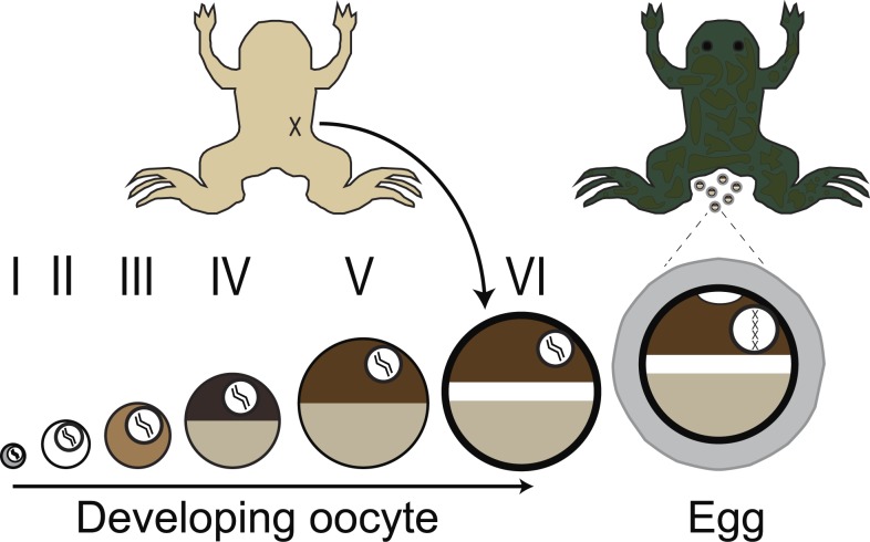
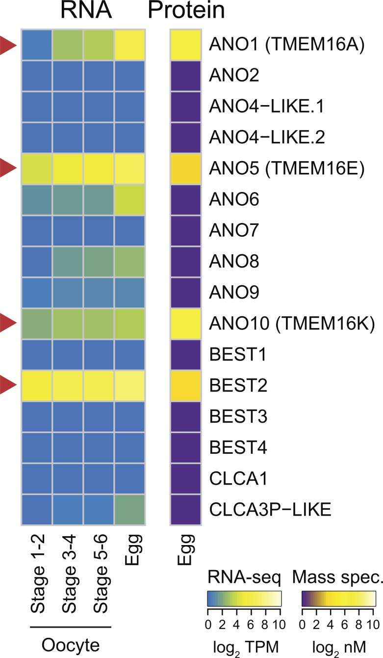
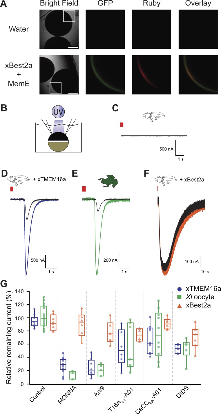
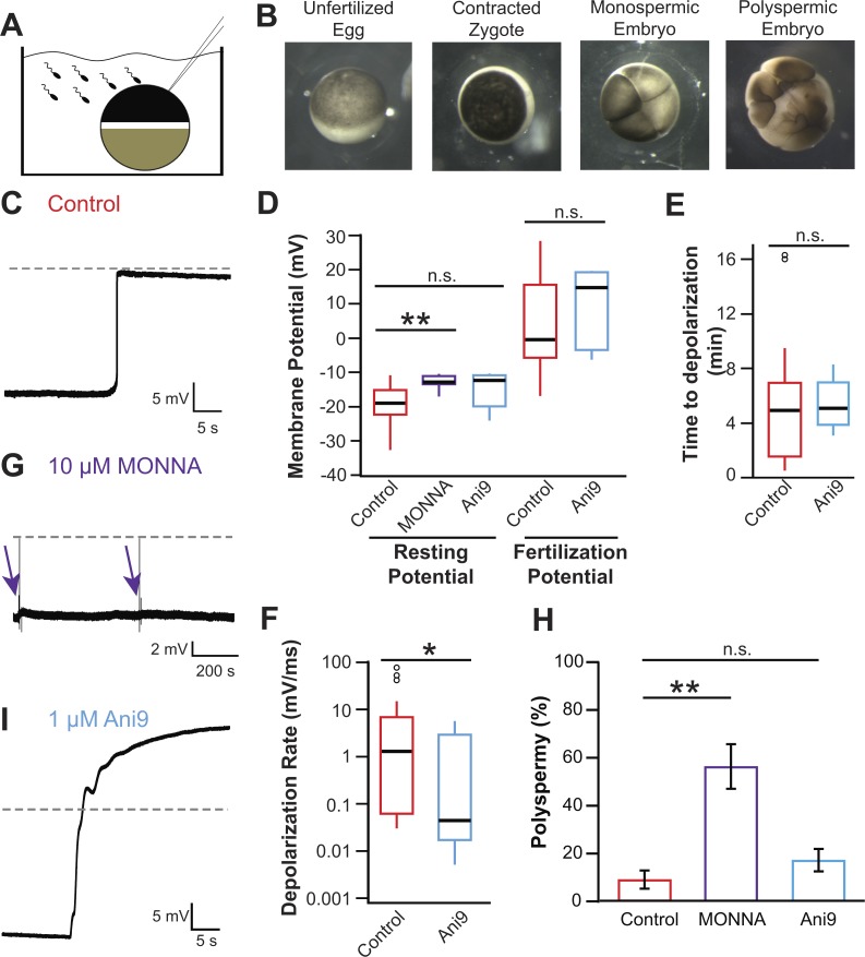
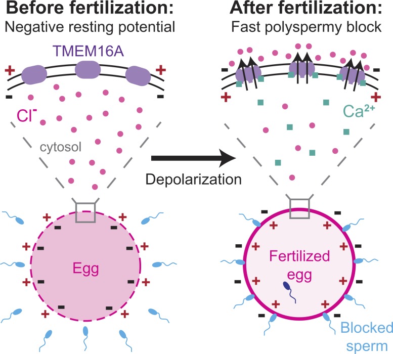
In externally fertilizing animals, such as sea urchins and frogs, prolonged depolarization of the egg immediately after fertilization inhibits the entry of additional sperm-a phenomenon known as the fast block to polyspermy. In the African clawed frog Xenopus laevis, this depolarization is driven by Ca2+-activated Cl- efflux. Although the prominent Ca2+-activated Cl- currents generated in immature X. laevis oocytes are mediated by X. laevis transmembrane protein 16a (xTMEM16A) channels, little is known about the channels that contribute to the fast block in mature eggs. Moreover, the gamete undergoes a gross transformation as it develops from an immature oocyte into a fertilization-competent egg. Here, we report the results of our approach to identify the Ca2+-activated Cl- channel that triggers the fast block. By querying published proteomic and RNA-sequencing data, we identify two Ca2+-activated Cl- channels expressed in fertilization-competent X. laevis eggs: xTMEM16A and X. laevis bestrophin 2A (xBEST2A). By exogenously expressing xTMEM16A and xBEST2A in axolotl cells lacking endogenous Ca2+-activated currents, we characterize the effect of inhibitors on currents mediated by these channels. None of the inhibitors tested block xBEST2A currents specifically. However, 2-(4-chloro-2-methylphenoxy)-N-[(2-methoxyphenyl)methylideneamino]-acetamide (Ani9) and N-((4-methoxy)-2-naphthyl)-5-nitroanthranilic acid (MONNA) each reduce xTMEM16A currents by more than 70% while only nominally inhibiting those generated by xBEST2A. Using whole-cell recordings during fertilization, we find that Ani9 and MONNA effectively diminish fertilization-evoked depolarizations. Additionally, these inhibitors lead to increased polyspermy in X. laevis embryos. These results indicate that fertilization activates TMEM16A channels in X. laevis eggs and induces the earliest known event triggered by fertilization: the fast block to polyspermy.


Figures

Figure 1.
Figure 1.
Schematic depiction of gamete development in female X. laevis. Immature oocytes, ranging from the youngest (stage I) to the most developed (stage VI), are located within the ovaries. These oocytes can be surgically removed from the abdomen of the frog (shown in ventral view at top left) and are commonly used by electrophysiologists. Upon hormonal induction, stage VI oocytes mature into fertilization-competent eggs, which are laid by the frog (shown in dorsal view at top right). Oocytes and eggs differ with respect to membrane-localized proteins as well as the structure of the cytoskeleton.
Figure 2.
Figure 2.
Expression of CaCCs in X. laevis oocytes and eggs. Heatmaps of expression levels of CaCCs at the developmental stages indicated. Right: Protein concentrations (from Wühr et al., 2014) as determined by mass spectrometry–based proteomics study (in log2 nanomolar). Left: Transcript levels (shown as transcripts per million [TPM]; from Session et al., 2016), as determined by RNA-seq–based transcriptome study. Arrowheads highlight CaCCs with proteins found in eggs.
Figure 3.
Figure 3.
MONNA and Ani9 inhibit TMEM16A-conducted Cl currents. (A) Representative bright-field and fluorescence images of axolotl oocytes expressing Ruby-tagged xBEST2a and enhanced GFP–tagged MemE (reporter of plasma membrane). Boxes denote portions included in fluorescence images. Bars, 750 µm. Overlay is of GFP and Ruby images. (B) Schematic of experimental design: UV photolysis to uncage IP3 while conducting TEVC. (C–F) Current recordings from oocytes of axolotls (C, D, and F) or X. laevis (E), after injection with a photolabile caged-IP3 analogue, with clamping at −80 mV. Axolotl oocytes expressed no transgene (C), xTMEM16A (D), or xBEST2A (F). Wild-type X. laevis oocytes expressing endogenous channels (E). Typical current traces before and after uncaging, during (colored) control treatment, and (black) in the presence of 10 µM MONNA. Red bar denotes the 250-ms duration of UV exposure. (G) Tukey box plot distributions of the averaged proportion of current remaining after application of the indicated inhibitors, in axolotl oocytes expressing xTMEM16A (n = 6–14) or xBEST2A (n = 6–8), and in X. laevis oocytes expressing endogenous channels (n = 5–16). The central line represents the median value and the box denotes the data spread from 25–75%, and the whiskers reflect 10–90%.
Figure 4.
Figure 4.
Fertilization activates TMEM16A to depolarize the egg. (A) Schematic depiction of experimental design showing whole-cell recordings made on X. laevis eggs during fertilization. (B) Images of an X. laevis egg before sperm addition (left), an egg ∼15 min after fertilization with animal pole contracted (center left), a monospermic embryo (center right), and a polyspermic embryo (right). (C, G, and I) Representative whole-cell recordings made during fertilization in control conditions (C), the presence of 10 µM MONNA (G), or the presence of 1 µM Ani9 (I). Dashed lines denote 0 mV, and arrows denote times at which sperm was applied to eggs in the presence of 10 µM MONNA. (D–F) Tukey box plot distributions of the resting and fertilization potentials in control conditions and with MONNA or Ani9 (D), the time between sperm application and depolarization in the absence and presence of Ani9 (E), and the depolarization rate in the absence and presence of Ani9 (F; n = 5–30, recorded over 2–16 experimental days per treatment). The central line represents the median value, and the box denotes the data spread from 25–75%, and the whiskers reflect 10–90%. (H) Proportion of polyspermic embryos out of total developed embryos in control, MONNA, and Ani9 (n = 3, recorded over three experiment days per treatment). n.s., P > 0.05; *, P < 0.05; **, P < 0.001.
Figure 5.
Figure 5.
Proposed model for fertilization signaled activation of TMEM16A. Before fertilization, X. laevis eggs have a negative resting potential, thereby electrically signaling to sperm that they can receive a male gamete. After fertilization, cytosolic Ca2+ increases to activate TMEM16Aa. An efflux of Cl then depolarizes the egg, and this change in membrane potential blocks supernumerary sperm from entering the fertilized egg.

Comment in

References

    1. Brunner J.D., Lim N.K., Schenck S., Duerst A., and Dutzler R.. 2014. X-ray structure of a calcium-activated TMEM16 lipid scramblase. Nature. 516:207–212. 10.1038/nature13984 - DOI - PubMed
    1. Bulley S., Neeb Z.P., Burris S.K., Bannister J.P., Thomas-Gatewood C.M., Jangsangthong W., and Jaggar J.H.. 2012. TMEM16A/ANO1 channels contribute to the myogenic response in cerebral arteries. Circ. Res. 111:1027–1036. 10.1161/CIRCRESAHA.112.277145 - DOI - PMC - PubMed
    1. Caputo A., Caci E., Ferrera L., Pedemonte N., Barsanti C., Sondo E., Pfeffer U., Ravazzolo R., Zegarra-Moran O., and Galietta L.J.. 2008. TMEM16A, a membrane protein associated with calcium-dependent chloride channel activity. Science. 322:590–594. 10.1126/science.1163518 - DOI - PubMed
    1. Cross N.L. 1981. Initiation of the activation potential by an increase in intracellular calcium in eggs of the frog, Rana pipiens. Dev. Biol. 85:380–384. 10.1016/0012-1606(81)90269-4 - DOI - PubMed
    1. Cross N.L., and Elinson R.P.. 1980. A fast block to polyspermy in frogs mediated by changes in the membrane potential. Dev. Biol. 75:187–198. 10.1016/0012-1606(80)90154-2 - DOI - PubMed

Publication types