. 2018 Aug;25(8):660-668.
doi: 10.1038/s41594-018-0089-6. Epub 2018 Jul 16.

Structural basis for the regulation of inositol trisphosphate receptors by Ca2+ and IP3

Affiliations

Structural basis for the regulation of inositol trisphosphate receptors by Ca2+ and IP3

Navid Paknejad et al. Nat Struct Mol Biol. 2018 Aug.

Erratum in

Abstract

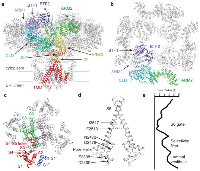
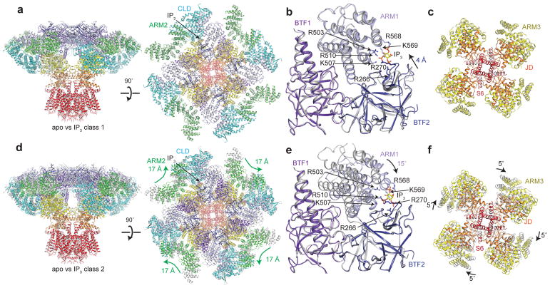
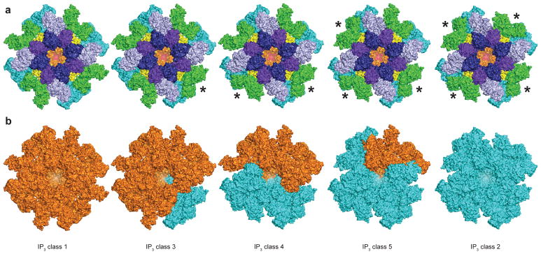
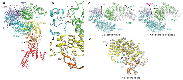
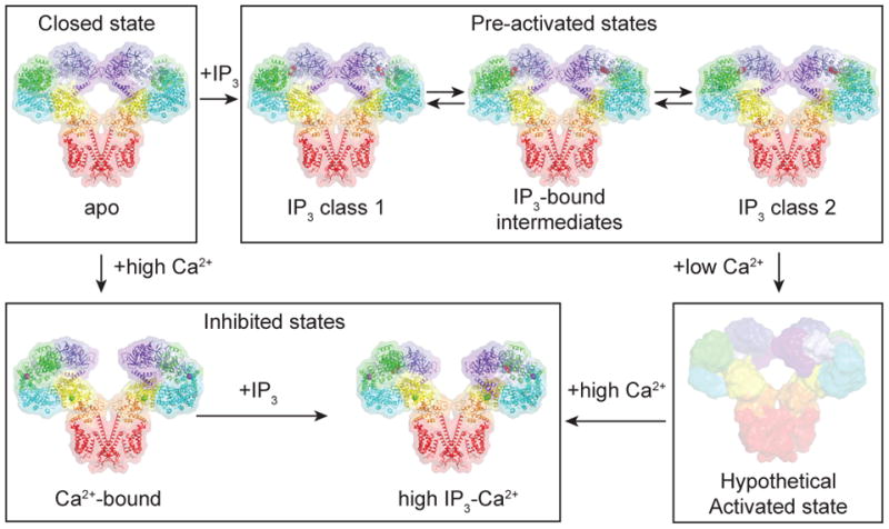
Inositol trisphosphate receptors (IP3Rs) are ubiquitous Ca2+-permeable channels that mediate release of Ca2+ from the endoplasmic reticulum, thereby regulating numerous processes including cell division, cell death, differentiation and fertilization. IP3Rs are jointly activated by inositol trisphosphate (IP3) and their permeant ion, Ca2+. At high concentrations, however, Ca2+ inhibits activity, ensuring precise spatiotemporal control over intracellular Ca2+. Despite extensive characterization of IP3R, the mechanisms through which these molecules control channel gating have remained elusive. Here, we present structures of full-length human type 3 IP3Rs in ligand-bound and ligand-free states. Multiple IP3-bound structures demonstrate that the large cytoplasmic domain provides a platform for propagation of long-range conformational changes to the ion-conduction gate. Structures in the presence of Ca2+ reveal two Ca2+-binding sites that induce the disruption of numerous interactions between subunits, thereby inhibiting IP3R. These structures thus provide a mechanistic basis for beginning to understand the regulation of IP3R.


Conflict of interest statement

Competing financial interest statement

The authors declare no competing interests.

Figures

Figure 1
Figure 1. Structure of human type 3 IP3R in a ligand-free state
a, Structure of hIP3R3 viewed in the plane of the membrane with the cytoplasmic domain at the top. A single subunit is colored according to domain with BTF1 in purple, BTF2 in blue, ARM1 in violet, the CLD in cyan, ARM2 in green, ARM3 in yellow, the JD in orange and the TMD in red. Gray lines represent the approximate position of the membrane. b, Structure of the cytoplasmic domain viewed from the cytoplasm. A single subunit is colored according to domain. ARM3 is removed for clarity. c, Structure of the transmembrane domain viewed from the cytoplasm. S1–S4 helices are colored red, S1′ and S1″ blue and S5, S6 and pore helix green. d–e, Structure of the ion conduction pathway viewed in the plane of the membrane and plot of pore radius. Residues comprising the luminal vestibule, the selectivity filter and S6 gate are highlighted. Front and rear subunits are removed for clarity.
Figure 2
Figure 2. IP3 binding site in two IP3-bound conformations
a, d, Superposition of (a) IP3 class 1 (colored by domain) and apo (grey) or (d) IP3 class 2 (colored by domain) and apo (grey) viewed from in the plane of the membrane (left) and the cytoplasm (right). b, e, Superposition of IP3-binding domain of (b) IP3 class 1 (colored by domain) and apo (grey) or (e) IP3 class 2 (colored by domain) and apo (grey) aligned by BTF1 and BTF2. IP3 and the side chains of IP3-coordinating residues are shown as sticks. c, f, Superposition of ARM3, the JD and S6 of (c) IP3 class 1 (colored by domain) and apo (grey) or (f) IP3 class 2 (colored by domain) and apo (grey) aligned by the TMD and viewed from the lumen.
Figure 3
Figure 3. Ensemble of IP3-bound conformations
a, Structures of IP3 class 1, IP3 class 32, IP3 class 4, IP3 class 5 and IP3 class 2 colored by domain. The ARM2 domain of subunits marked with an * adopt a class 2-like conformation. b, Structures of IP3 class 1, IP3 class 3, IP3 class 4, IP3 class 5 and IP3 class 2. Domains adopting class 1-like domains are colored orange and domains adopting class 2-like conformations are colored cyan.
Figure 4
Figure 4. Ca2+ binding sites in Ca2+-bound hIP3R3
a, Monomeric structure of Ca2+-bound hIP3R3 colored by domain viewed in the plane of the membrane. Ca2+ and Zn2+ ions are shown as spheres. b, CD Ca2+-binding site at the CLD-ARM2 interface. Residues coordinating the Ca2+ are shown as sticks and the CD Ca2+ ion is shown as a magenta sphere. c, Superposition of the CLD and ARM2 of Ca2+-bound (colored by domain) and apo (grey, left) or Ca2+-bound (colored by domain) and IP3 class 2 (grey, right) aligned by the CLD. d, JD Ca2+-binding site at the ARM3-JD interface. Residues coordinating the Ca2+ are shown as sticks and the Ca2+ ion is shown as a green sphere. e, Superposition of ARM3 and the JD of Ca2+-bound (colored by domain) and apo (grey) aligned by the JD.
Figure 5
Figure 5. Ca2+ binding disrupts CD intra- and inter-subunit interactions
a–b, Cytoplasmic domain of (a) apo and (b) Ca2+-bound colored by domain, viewed from the cytoplasm. Spheres represent the Cα position of Trp168 of BTF1 and Lys426 of BTF2 (pink) and Pro140 of BTF1 and Ala1291 of ARM2 (brown). c–d, Structure of (c) apo and (d) Ca2+-bound colored by domain, viewed in the plane of the membrane. Front and rear subunits are removed for clarity. Ca2+ and Zn2+ ions are shown as spheres. Grey lines represent the approximate position of the membrane.
Figure 6
Figure 6. IP3-induced conformational changes
a, High IP3-Ca2+ structure colored by distance of Cα deviation from the Ca2+-bound structure viewed in the plane of the membrane. b, IP3 class 2 colored by distance of Cα deviation from the apo structure viewed in the plane of the membrane. c, Cytoplasmic domain of high IP3-Ca2+ structure colored by distance of Cα deviation from the Ca2+-bound structure viewed from the cytoplasm. d, Cytoplasmic domain of IP3 class 2 colored by distance of Cα deviation from the apo structure viewed from the cytoplasm. IP3 and Ca2+ ions are shown as spheres.
Figure 7
Figure 7. Model for IP3R regulation by IP3 and Ca2+
In the absence of Ca2+ and IP3, IP3R adopts a closed state. In the presence of IP3, IP3R adopts one of an ensemble of pre-activated states that are in equilibrium. In the presence of high Ca2+, the BTF ring is dissociated and the channel adopts an inhibited state. In the presence of high Ca2+ and IP3, the BTF ring is dissociated and the channel adopts an inhibited state. In the presence of low Ca2+ and IP3, the channel adopts a hypothetical activated state in which the S6 gate is opened.

References

    1. Burgess GM, Mckinney JS, Fabiatoq A, Leslie BA, Putney JW. Calcium Pools in Saponin-permeabilized Guinea Pig Hepatocytes. J Biol Chem. 1983;258:35336–153345. - PubMed
    1. Ehrlich BE, Watras J. Inositol 1,4,5-trisphosphate activates a channel from smooth muscle sarcoplasmic reticulum. Nature. 1988;336:583–586. - PubMed
    1. Furuichi T, et al. Primary structure and functional expression of the inositol 1,4,5-trisphosphate-binding protein P400. Nature. 1989;342:32–38. - PubMed
    1. Mignery GA, Südhof TC, Takei K, De Camilli P. Putative receptor for inositol 1,4,5-trisphosphate similar to ryanodine receptor. Nature. 1989;342:192–5. - PubMed
    1. Streb H, Irvine RF, Berridge MJ, Schulz I. Release of Ca2+from a nonmitochondrial intracellular store in pancreatic acinar cells by inositol-1,4,5-trisphosphate. Nature. 1983;306:67–69. - PubMed

Publication types

MeSH terms