. 2018 Jul 16;8(1):10705.
doi: 10.1038/s41598-018-29058-6.

Structural determinants of specificity and regulation of activity in the allosteric loop network of human KLK8/neuropsin

Affiliations

Structural determinants of specificity and regulation of activity in the allosteric loop network of human KLK8/neuropsin

Mekdes Debela et al. Sci Rep. .

Abstract

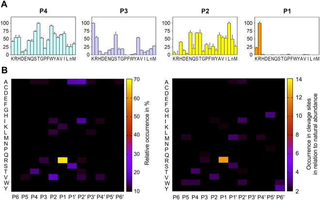
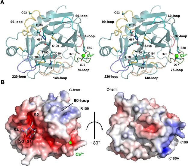
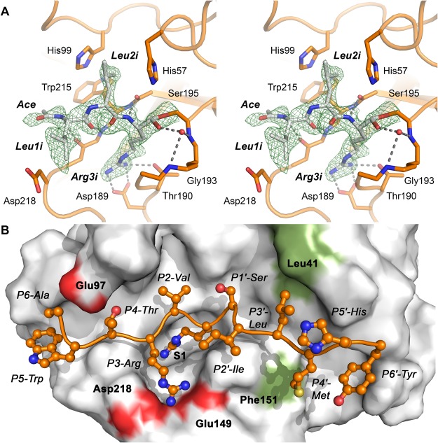
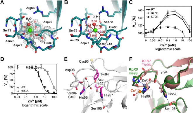
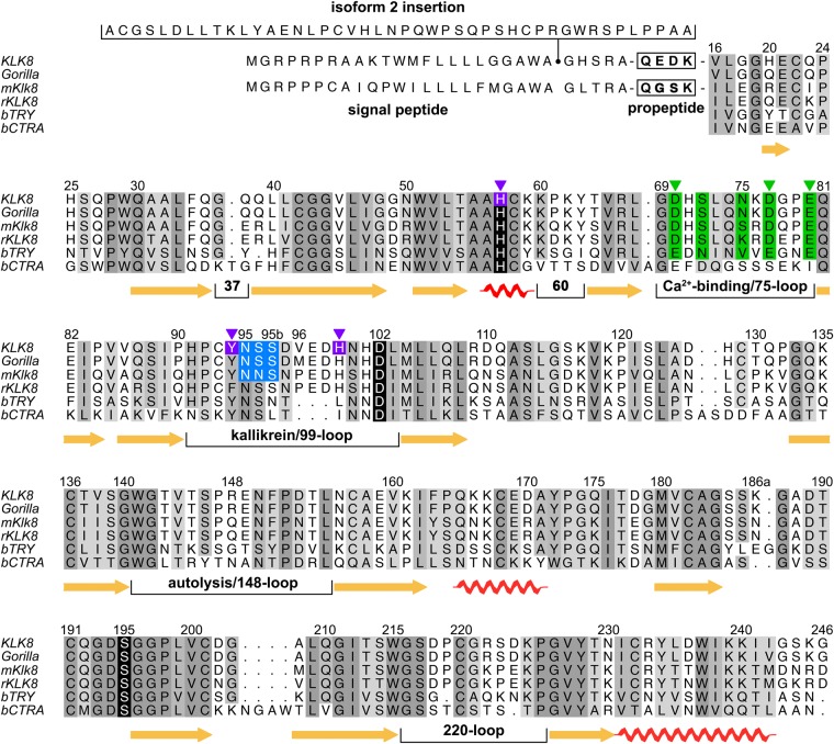
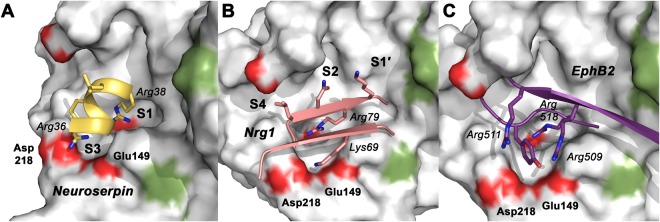
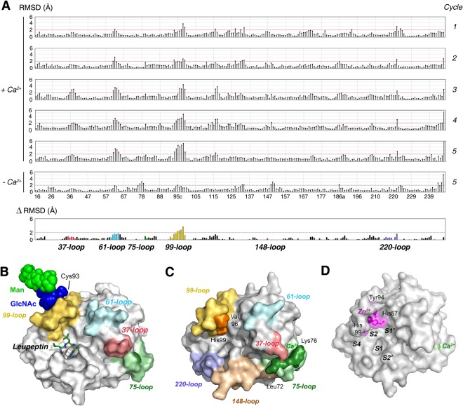
Human KLK8/neuropsin, a kallikrein-related serine peptidase, is mostly expressed in skin and the hippocampus regions of the brain, where it regulates memory formation by synaptic remodeling. Substrate profiles of recombinant KLK8 were analyzed with positional scanning using fluorogenic tetrapeptides and the proteomic PICS approach, which revealed the prime side specificity. Enzyme kinetics with optimized substrates showed stimulation by Ca2+ and inhibition by Zn2+, which are physiological regulators. Crystal structures of KLK8 with a ligand-free active site and with the inhibitor leupeptin explain the subsite specificity and display Ca2+ bound to the 75-loop. The variants D70K and H99A confirmed the antagonistic role of the cation binding sites. Molecular docking and dynamics calculations provided insights in substrate binding and the dual regulation of activity by Ca2+ and Zn2+, which are important in neuron and skin physiology. Both cations participate in the allosteric surface loop network present in related serine proteases. A comparison of the positional scanning data with substrates from brain suggests an adaptive recognition by KLK8, based on the tertiary structures of its targets. These combined findings provide a comprehensive picture of the molecular mechanisms underlying the enzyme activity of KLK8.


Conflict of interest statement

The authors declare no competing interests.

Figures

Figure 1
Figure 1
Specificity profiling with a positional scanning and proteomic identification of cleavage sites. (A) Peptide substrate specificity of KLK8 measured by positional scanning for peptide positions P4 to P1. The height of the columns represents substrate cleavage rates by released ACC, while the x-axis indicates the amino acids in one-letter code, whereby n represents norleucine, which substitutes Cys. (B) PICS heat maps with amino acids listed in alphabetical order along the y-axis and peptide positions run from P6 to P6′ on the x-axis. The left panel displays cleavages in 73 substrates and the right panel contains corrected values for the natural abundance of each residue. Both PICS and PSSCL agree in a moderate preference for Thr and Trp in P4 and for basic residues in P3, such as Lys or Arg. Aliphatic residues Leu, Val, and Ile are found by both methods in P2, whereby Val dominates in the corrected PICS, while PSSCL ranks the polar Asn, Ser, and Thr relatively high. The major primary specificity for Arg over Lys in P1 is found by both methods, albeit stronger in PICS. The corrected PICS data show a preference for Ser and Met in P1′, for Ile and Trp in P2′, and no distinct specificity for P3′ and P4′. By contrast, S5′ and S6′ subsites are moderately specific for His and Tyr.
Figure 2
Figure 2
Overall structure of KLK8-Ca. (A) Ribbon stereo plot of KLK8-Ca shown in standard orientation. The side chains of the catalytic triad, the activating salt bridge Val16-Asp194, the major specificity-determining Asp189 and Thr190, as well as the six disulfide bridges and Cys93 are shown as sticks (carbon teal, nitrogen blue, oxygen red, and sulfur yellow). Also, the three major ligands of Ca2+ (green sphere) are depicted, namely Asp70, Asp77 and Glu80. Functionally important loops are displayed in different colors with labels. (B) Surface model of KLK8 with inhibitor and Ca2+. The left panel shows KLK8 in standard orientation with a stick model of leupeptin, occupying the subsites S4 to S1. The electrostatic surface potential is depicted red for negative and blue for positive potential in the range −10 e/kBT to +10 e/kBT. The right panel shows the KLK8 backside, rotated by 180°. The 60-loop and the area around Arg109 correspond to the thrombin anion binding exosite I, while the positive patch at the back around Lys166 and Lys186A has no counterpart.
Figure 3
Figure 3
Leupeptin inhibitor and ideal substrate binding to the active site. (A) Stereo model of leupeptin in KLK8-leup, with the most relevant interacting residues displayed as sticks, e.g. the covalently linked Ser195. The electron density is a 2Fo-Fc map shown as green grid, contoured at 1.0 σ, hydrogen bonds are represented by grey dots. A water molecule (red sphere) occupies the oxyanion hole at the NH groups of Gly193 and Ser195 in this chain. (B) Molecular docking of an ideal substrate to KLK8 according to PICS profiling for residues P6 to P6′. Binding of residues P4 to P4′ agrees well with the specificity profiles. Moreover, the calculated interactions of P4-Thr with Glu97 and P3-Arg with Glu149 and Asp218 (red patches) can explain the preference of the S4 subsite for Thr over Trp, and the unique S3 preference for basic side chains. Binding of the hydrophobic residues P3′-Leu and P4′-Met may depend on the subsite shaping residues Leu41 and Phe151 (green patches).
Figure 4
Figure 4
The Ca2+ and Zn2+ binding loops of KLK8 as regulatory modules. (A) The 75-loop of KLK8-Ca with Ca2+ ligands surrounded by the 2Fo-Fc electron density map, contoured at 1.0 σ. Ca2+ is depicted in green with electron density contoured at 3.0 σ, while the water ligand is shown as red sphere. (B) Ligand bonds to Ca2+ are shown as red dots and relevant hydrogen bonds as black dots, including distances in Å. The excess charge of three carboxylates is compensated by Arg66 near Asp70 and hydrogen bonds of Asp77 and Glu80 with backbone N-H groups. (C) The Ca2+ stimulation of the KLK8 activity is maximal in the range 100 µM to about 1 mM. In the D70K variant no significant stimulatory effect of Ca2+ was observed. (D) The mutant His99Ala confirmed the inhibitory Zn2+ site with a 13-fold higher IC50, i.e. 47 µM versus 3.6 µM of wild-type KLK8. (E) In the 99-loop, Cys93 can be excluded as potential Zn2+ ligand, since Zn2+ inhibition of the Cys93Ser variant was unchanged compared to the wild-type. A water molecule (grey) between His99 and Tyr94 in KLK8-leup is in a suitable position to bind Zn2+. The variant Tyr94Phe has a 2-fold higher IC50, suggesting a minor role as Zn2+ ligand for Tyr94. Hydrogen bonds are shown as grey dots. (F) Two complex structures of KLK5 and KLK7 with Zn2+ and Cu2+ feature ligands for cations, such as the carbonyl O of residue 96 and Tyr94. The KLK7-His99Ala mutant nearly abolished the non-competitive Zn2+ inhibition, as for KLK8. This inhibition type can be explained by Zn2+ binding to the His57 side chain, which has to switch from the catalytic triad. A rotation of His99 would bring both ligands in ideal distances to Zn2+ .
Figure 5
Figure 5
Structure-based sequence alignment of KLK8. Other species are gorilla (Gorilla gorilla), mouse (Mus musculus) and rat (Rattus norvegicus) (mKlk8 and rKLK8), including bovine trypsin (bTRY) and chymotrypsinogen A (bCTRA) as numbering standard. Signal peptides of isoforms precede the propeptide, whereby isoform 2 with a 45 residue insertion is supposed to be unique for humans. Human and chimpanzee KLK8 are identical, whereas the gorilla has three exchanges. Mice share 73% identical residues in KLK8 with humans, similar to rats with about 71%. β-sheets, α-helices, and the short 310-helix (residues 56–59) of KLK8 are indicated by arrows and screws, respectively. Relevant surface loops are labeled according to the central residue. Darker shades of grey represent higher conservation, while the catalytic triad residues are depicted with black background. Ca2+-ligands are displayed with green background and Zn2+ ligands are shown with magenta background, triangles indicate interacting side chains. The sequons of the glycosylation site at Asn95 are highlighted in blue.
Figure 6
Figure 6
Models of natural protein substrates bound to the active site of KLK8. The molecular surface of KLK8 is shown with red patches of acidic residues and green for hydrophobic ones. (A) Human neuroserpin, a serine protease inhibitor from the hippocampus, is cleaved at Arg38 as P1 residue, which is located in the last turn of an α-helix, as observed in the crystal structure (PDB 3FGQ). Apparently, Arg36, Leu37 and Ala39 are suitable P3, P2 and P1′ residues in accordance with our profiling results. Proper binding and turnover by KLK8 requires a transition of the helical turn into a more linear, extended conformation. (B) The synaptic regulator neuregulin 1 (NRG1) possesses only P1-Arg79 and P2-Asn78 as KLK8 specific substrate residues at its cleavage site. According to a structure-based homology model, created by SWISS-MODEL they belong to a β-strand, while the Leu77 side-chain might occupy the S4 instead of the S3 subsite. However, an adjacent β-strand contains Lys69, which is located close to Glu149 and Asp218, substitute the preferred basic P3 side-chain. (C) Similarly, a model from SWISS-MODEL of the neuronal KLK8 substrate ephrin type-B receptor 2 (EphB2), exhibits an arrangement of two strands, with P1-Arg518 as only specific residue for cleavage by KLK8. Interestingly, Arg509 and Arg511 might be ideally positioned for electrostatic interaction with Glu149 and Asp218.
Figure 7
Figure 7
Molecular dynamics and the allosteric loop network of KLK8. (A) Plots of root mean square deviations (RMSD) in Å of KLK8 residue coordinates for five sequential MD production cycles at 298 K after the warm-up cycles, with respect to the final warm-up model. Data are depicted Ca2+ bound to the 75-loop and the last cycle without Ca2+ (chymotrypsinogen numbering). The significance level 2 Å (red line) is MOE default. For better visualization of loop motions, the RMSD difference (∆) of the models from cycle 5 [(+Ca2+) − (−Ca2+)] was calculated. Whereas the Ca2+ bound 75-loop seems more stationary, the 37-, 61-, and mainly the 99-loop exhibit large motions. This finding indicates an allosteric communication line between these loops. (B) Model from MD calculations of KLK8 shown as surface with colored loops with a core glycan linked to Asn95, consisting of two GlcNAc (blue spheres) and three mannoses (light green). Ca2+ (green) is bound to the 75-loop and leupeptin from the crystal structure is overlayed as stick model at subsites S4 to S1, in order to emphasize the wide open 99-loop (yellow) conformation, which seems to be facilitated by the presence of the glycan. The glycan may prevent cleavage in the 99-loop and disulfide formation of Cys93. (C) In MD calculations of glycan-free KLK8 were similar as for glycosylated KLK8. Upon removal of Ca2+, the 75-loop closes the substrate binding cleft in the prime side near the 37-loop. The concomitant closure of the S2 pocket by Val96 and His99 (orange) in the 99-loop might hamper binding of small substrates. The labeled loops around the active site represent the allosteric network of various trypsin-like serine proteases, which may regulate substrate access. (D) Zn2+-binding would similarly close the S2 pocket and inactivate KLK8 by rotating the His57 side chain away from the catalytic triad. Such a conformation was observed in the structure of rat tonin (1TON).

References

    1. Suzuki J, et al. Ontogeny of neuropsin mRNA expression in the mouse brain. Neuroscience Research. 1995;23:345–351. doi: 10.1016/0168-0102(95)00960-2. - DOI - PubMed
    1. Yoshida S, Taniguchi M, Hirata A, Shiosaka S. Sequence analysis and expression of human neuropsin cDNA and gene. Gene. 1998;213:9–16. doi: 10.1016/S0378-1119(98)00232-7. - DOI - PubMed
    1. Kishi T, Grass L, Soosaipillai A, Shimizu-Okabe C, Diamandis EP. Human Kallikrein 8: Immunoassay Development and Identification in Tissue Extracts and Biological Fluids. Clin Chem. 2003;49:87–96. doi: 10.1373/49.1.87. - DOI - PubMed
    1. Shaw JLV, Diamandis EP. Distribution of 15 Human Kallikreins in Tissues and Biological Fluids. Clin Chem. 2007;53:1423–1432. doi: 10.1373/clinchem.2007.088104. - DOI - PubMed
    1. Rawlings ND, Barrett AJ, Bateman A. MEROPS: the database of proteolytic enzymes, their substrates and inhibitors. Nucleic Acids Res. 2012;40:D343–350. doi: 10.1093/nar/gkr987. - DOI - PMC - PubMed

Publication types

MeSH terms